Quick Details
color doppler ultrasound pregnancy ultrasound color doppler pregnancy portable color doppler ultrasound machine
Packaging & Delivery
| Packaging detail:Standard export package Delivery detail:within 7-10 workdays after receipt of payment |
Specifications
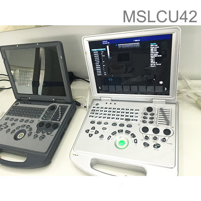
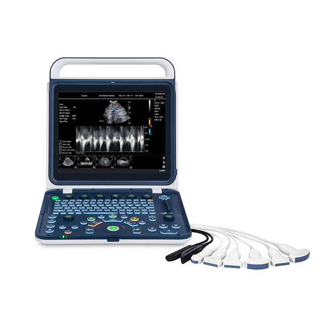
Portable color doppler ultrasound 80 element probe AMCU42
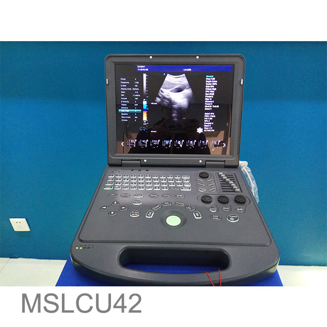
Portable color doppler ultrasound Main features:
With high precision digital beam forming and Doppler ultrasonic imaging technology, Pro incorporated the latest image processing technologies such as THI, speckle reduction, multi-beam parallel processing and efficient full-digital image management system is easy to acquire better image. Special measurement software packages, flexible configuration and ergonomical design greatly increase operators clinical diagnosis accruacy and analysis efficiency.  color doppler ultrasound pregnancy Application Abdomen, OB&GYN, cardiology, vascular and small parts, urology, musculoskeletal, pediatrics and etc Portable color doppler ultrasound Displaying mode B, 2B, 4B, left&right, B|M, B|D, PW, M, B mode, part zoom, B|C|D, B|C|M, B|C, duplex, PW, CFM, CPA
  Portable color doppler ultrasound Signal processing Full-digital beam forming, dynamic filter, orthogonal demodulating, space-time filter, dynamic real-time receiving focusing, RDA, DRA, spectral processing, CFM processing Portable color doppler ultrasound Image processing THI, speckle-reduction, color coder, frame averaging, micro- angle adjustment, wall filter, 256 grey scale, scanning angle/width control, composit processing of tissue and blood flow image
 Portable color doppler ultrasound 80 element probe AMCU42 video
Portable color doppler ultrasound General measurement B mode: distance, angle, perimeter and area (ellipse method, Trace method), volume, histogram, cross-section diagram M mode: cardiac rate, time, distance, speed. color doppler ultrasound pregnancy Measurement & report packages GYN(four edition for GA calculation), cardiac, vascular, urology, andriatrics, peripheral vascular, multiple births, orthopedic surgery and etc. -Storage function Probe parameter, image, cine loop, measurement data and report -Cine loop Operated by automatically and manually, speed optional, searching cine loop, forward/ backward cine loop – Input/output interface VGA, network, USB, VIDEO, parallet communication port, serial communication port Portable color doppler ultrasound Standard configuration Main unit, 3.5MHz convex probe, 15″ LCD monitor, one probe connector, USB port DICOM3.0 color doppler ultrasound pregnancy Optional configuration 7.5Mhz(2.0-10.0Mhz) linear probe 7.0MHz(2.0-10.0Mhz) trans-vaginal probe 2.0MHz(2.0-10.0Mhz) Microconvex probe Thermal printer laser printer Trolley Appearance Weight: 7.3kgs Display:15 inch LCD Monitor Power Requirements Power requirements : AC (AC) 220V ± 22V 50Hz ± 1Hz Input power : 350VA Scanning :Electronic linear array, electronic convex array Display Mode: Black and white image : B, 2B, 4B, left B | M, B | D, PW, M, B partial enlarged mode Bloodstream image : Spectrum : B / D, B / C / D CFM: B | C | D, B | C | M, B | C pairs in real time , PW, CFM, CPA Color flow image adjustment parameters : a Doppler frequency , sampling frame position and size , baseline color gain, the deflection angle , the wall filter, the cumulative frequency. Display depth:Continuously adjustable Focus: Electronic focus + Acoustic lens focus Launch with a single focus, bifocal , trifocal and four focus work Receiving continuous dynamic focusing Signal processing / Doppler sound: Dynamic Filtering Quadrature demodulator The total gain adjustment Composite Gain Technology: 8-segment TGC and D-AGC B -, C -, D- , respectively, the total gain is adjustable Low pass filter Subsample Emitter voltage regulation Doppler tone adjustable stereo output Image processing: Logarithmic conversion and dynamic range compression Temporal filtering Spatial filtering Frame correlation Edge Enhancement Grayscale conversion B | M or M -type scanning speed Line density control Scan angle / width control Image optimization Flip the image around Image upside down Flip the image in black and white Multi-angle image rotation Choose a color grayscale inversion Article Tissue blood flow images and image processing complex Black and white image resolution 1024 × 768 × 8bit (256 gray levels ) Color Image Resolution 1024 × 768 8bit × 8bit × 8bit color coding The basic measurement and calculation functions: B -mode basic measurements: distance, angle, perimeter and area ( ellipse method , track method ) , volume histograms , cross section M -mode basic measurements: heart rate, time , distance, speed Obstetric measurement and calculation functions: Obstetric report Measurement of amniotic fluid index calculation (AFI) Calculated ratio (BPD / OFD, FL / AC, FL / BPD, HC / AC) Estimation of fetal weight By the LMP, BBT projections gestational age and expected date function Fetal physiology score (Fetal Biophysical Score) Fetal growth curve Andrology measurement and calculation functions: Prostate , testicular and other measurements to calculate Prostate -specific antigen predicted PPSA, prostate specific antigen density calculated PSAD Gynecological measurement and calculation functions: Uterus, left ovary , right ovary , left follicle , the follicle and other measurements to calculate the right Urology measurement and calculation functions: Left kidney, kidney , bladder, residual urine volume (Residual urine vol) and other measurements to calculate Peripheral vascular measurement and calculation functions: Area stenosis , diameter stenosis measurement and calculation Small organ measurement and calculation functions: Thyroid, breast , and other measurements to calculate mass Orthopaedic measurement and calculation functions: Left hip , right hip measurement Cardiac function measurement and calculation: Heart measurement software package – providing heart rate, speed, left ventricle , aortic , mitral , ventricular ( right / left ventricle ) and other analytical measurement methods Area stenosis percentage (% Area Sten), percentage diameter stenosis (% Diam Sten) Body surface area BSA  Display information: Probe information , display mode, the depth of focus , the dynamic range of standard body , the probe position mark , sound power , patient information, medical institution name, the measurement value, time and date , the scale , the scanning direction , gamma curve , the probe current work Frequency, frame rate , B mode overall gain , C mode overall gain , D model of the total gain , menus, comments, gray belt, puncture guide line , PRF, wall filter , blood -related , cumulative frequency , TI heat index , MI machinery index Storage function: Probe parameter storage Image Storage Cine storage Measurement results are stored Report storage Grayscale :256 gray Interface Language:Optional English

