01 What is an ultrasound examination?
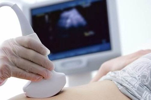
Talking about what ultrasound is, we must first understand what ultrasound is. Ultrasonic wave is a kind of sound wave, which belongs to mechanical wave. Sound waves with frequencies above the upper limit of what the human ear can hear (20,000 Hz, 20 KHZ) are ultrasound, while medical ultrasound frequencies typically range from 2 to 13 million Hz (2-13 MHZ). The imaging principle of ultrasound examination is: Due to the density of human organs and the difference in the speed of sound wave propagation, ultrasound will be reflected in different degrees, the probe receives the ultrasound reflected by different organs and is processed by the computer to form ultrasonic images, thus presenting the ultrasonography of each organ of the human body, and the sonographer analyzes these ultrasonography to achieve the purpose of diagnosis and treatment of diseases.
02 Is ultrasound harmful to human body?
A large number of studies and practical applications have proved that ultrasound examination is safe for the human body, and we need not feel anxious about it. From the principle analysis, ultrasound is the transmission of mechanical vibration in the medium, when it spreads in the biological medium and the irradiation dose exceeds a certain threshold, it will have a functional or structural impact on the biological medium, which is the biological effect of ultrasound. According to its mechanism of action, it can be divided into: mechanical effect, thixotropic effect, thermal effect, acoustic flow effect, cavitation effect, etc., and its adverse effects mainly depend on the size of the dose and the length of inspection time. However, we can rest assured that the current ultrasonic diagnostic instrument factory are in strict compliance with the United States FDA and China CFDA standards, the dose is within the safe range, as long as the reasonable control of the inspection time, ultrasound inspection has no harm to the human body. In addition, the Royal College of Obstetricians and Gynaecologists recommends that at least four prenatal ultrasounds should be performed between implantation and birth, which is enough to prove that ultrasounds are recognized worldwide as safe and can be performed with complete confidence, even in fetuses.
03 Why is it sometimes necessary before examination "Empty stomach", "full urine", "urination"?
Whether it is "fasting", "holding urine", or "urinating", it is to avoid other organs in the abdomen to interfere with the organs we need to examine.
For some organ examination, such as liver, bile, pancreas, spleen, kidney blood vessels, abdominal vessels, etc., an empty stomach is required before examination. Because the human body after eating, the gastrointestinal tract will produce gas, and ultrasound is "afraid" of gas. When ultrasound encounters gas, due to the large difference in the conductivity of gas and human tissues, most of the ultrasound is reflected, so the organs behind the gas can not be displayed. However, many organs in the abdomen are located near or behind the gastrointestinal tract, so an empty stomach is needed to avoid the impact of gas in the gastrointestinal tract on image quality. On the other hand, after eating, the bile in the gallbladder will be discharged to help digestion, the gallbladder will shrink, and even cannot be seen clearly, and the structure and abnormal changes in it will naturally be invisible. Therefore, before the examination of the liver, bile, pancreas, spleen, abdominal large blood vessels, kidney vessels, adults should be fasting for more than 8 hours, and children should be fasting for at least 4 hours.
When performing ultrasound examinations of the urinary system and gynecology (transabdominal), it is necessary to fill the bladder (hold urine) in order to show the relevant organs more clearly. This is because there is a bowel in front of the bladder, there is often gas interference, when we hold urine to fill the bladder, it will naturally push the intestine "away", you can make the bladder show clearly. At the same time, the bladder in the full state can more clearly show the bladder and bladder wall lesions. It's like a bag. When it's deflated, we can't see what's inside, but when we hold it open, we can see. Other organs, such as the prostate, uterus, and appendices, require a full bladder as a transparent window for better exploration. Therefore, for these examination items that need to hold urine, usually drink plain water and do not urinate 1-2 hours before the examination, and then check when there is a more obvious intention to urinate.
The gynecological ultrasound we mentioned above is an ultrasound examination through the abdominal wall, and it is necessary to hold urine before the examination. At the same time, there is another gynecologic ultrasound examination, that is, transvaginal gynecologic ultrasound (commonly known as "Yin ultrasound"), which requires urine before the examination. This is because transvaginal ultrasound is a probe placed in the woman's vagina, showing the uterus and the two appendages up, and the bladder is located just below the front of the uterine appendages, once it fills up, it will push the uterus and the two appendages back, making them away from our probe, resulting in poor imaging results. In addition, transvaginal ultrasound often requires pressure exploration, will also stimulate the bladder, if the bladder is full at this time, the patient will have a more obvious discomfort, may cause missed diagnosis.
04 Why the sticky stuff?
When doing ultrasound examination, the transparent liquid applied by the doctor is a coupling agent, which is a water-based polymer gel preparation, which can make the probe and our human body seamlessly connected, prevent the air from affecting the conduction of ultrasonic waves, and greatly improve the quality of ultrasonic imaging. Moreover, it has a certain lubricating effect, making the probe more smooth when sliding on the patient's body surface, which can save the doctor's strength and significantly reduce the patient's discomfort. This liquid is non-toxic, tasteless, non-irritating, rarely causes allergic reactions, and easy to clean, dry fast, check with a soft paper towel or towel can be wiped clean, or clean with water.
05 Doctor, wasn't my exam a "color ultrasound"?
Why are you looking at images in "black and white"
First of all, you need to understand that color ultrasound is not a color TV in our homes. Clinically, color ultrasound refers to color Doppler ultrasound, which is formed by superimposing the signal of blood flow onto the two-dimensional image of B-ultrasound (B-type ultrasound) after color coding. Here, the "color" reflects the blood flow situation, when we turn on the color Doppler function, the image will appear red or blue blood flow signal. This is an important function in our ultrasound examination process, which can reflect the blood flow of our normal organs and show the blood supply of the lesion site. The two-dimensional image of ultrasound uses different gray levels to represent different echoes of organs and lesions, so it looks "black and white". For example, the image below, the left is a two-dimensional image, it mainly reflects the anatomy of human tissue, looks "black and white", but when superimposed on the red, blue color blood flow signal, it becomes the right color "color ultrasound".
Left: "Black and white" ultrasound Right: "Color" ultrasound
06 Everyone knows that the heart is an extremely important organ.
So what do you need to know about cardiac ultrasound?
Cardiac echocardiography is a non-invasive examination using ultrasound technology to dynamically observe the size, shape, structure, valve, hemodynamics and cardiac function of the heart. It has important diagnostic value for congenital heart disease and heart disease, valvular disease and cardiomyopathy affected by acquired factors. Before doing this examination, adults do not need to empty stomach, nor do they need other special preparations, pay attention to suspending the use of drugs that affect heart function (such as digitalis, etc.), wear loose clothes to facilitate the examination. When children do cardiac ultrasound, because the crying of children will seriously affect the doctor's evaluation of the heart blood flow, children under the age of 3 are generally recommended to be sedating after the examination with the assistance of pediatricians. For children over 3 years of age, sedation can be determined according to the condition of the child. For children with severe crying and unable to cooperate with the examination, it is recommended to conduct an examination after sedation. For more cooperative children, you can consider direct examination accompanied by parents.
Post time: Aug-30-2023


