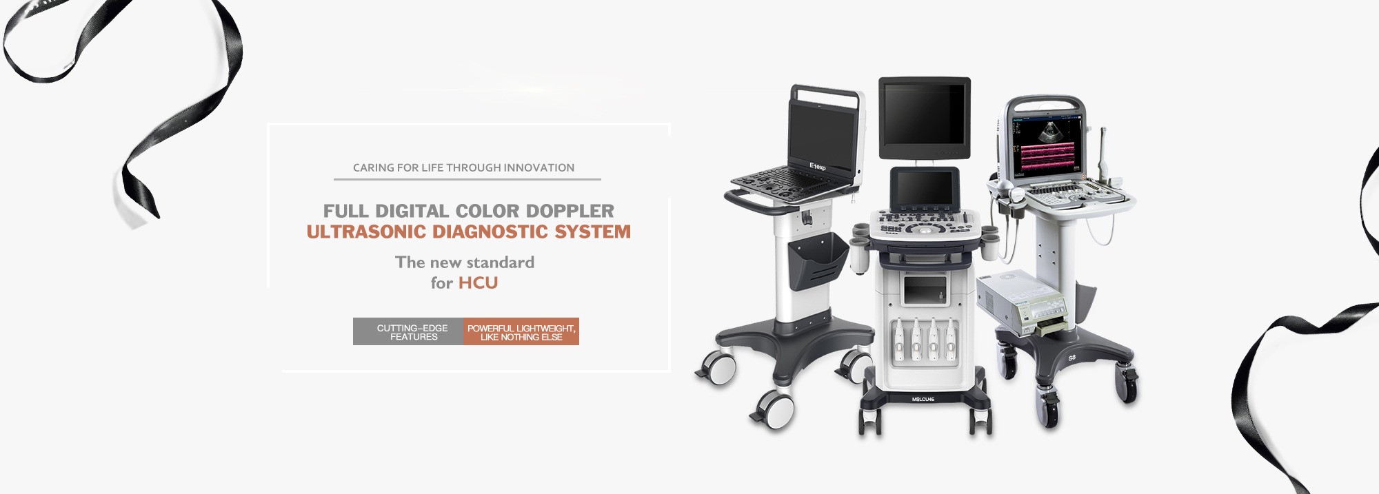
The economic benefit of sheep farm is directly related to the breeding characteristics of sheep. Veterinary ultrasound plays a very important role in the diagnosis of pregnancy of female animals. The pregnancy of the ewe can be determined by ultrasound.
The breeder/veterinarian can scientifically raise the pregnant ewes by means of grouping and individual shed feeding through the analysis of ultrasonic test results, so as to improve the nutritional management level of pregnant ewes and increase the lambing rate.
At this stage, for the ewe pregnancy inspection method, it is more commonly used to use animal B-ultrasound machine.
Veterinary B-ultrasound is usually used in animal pregnancy diagnosis, disease diagnosis, litter size estimation, identification of stillbirth, etc. It has the advantages of rapid examination and obvious results. Compared with the traditional detection methods in the past, veterinary ultrasound greatly simplifies the inspection process, reduces the inspection cost, and helps the breeder/veterinarian to quickly find the problem and adopt the response plan faster, such as: rapid group sorting.
What is B-ultrasound?
B-ultrasound is a high-tech means to observe the living body without any damage or stimulation, and has become a beneficial assistant for veterinary diagnostic activities and a necessary monitoring instrument for scientific research such as living egg collection and embryo transfer.
Domestic sheep are mainly divided into two categories: sheep and goats.
(1)Sheep breed
China's sheep breed resources are rich, product types are diverse. There are 51 sheep breeds of different production types, of which the fine sheep breeds account for 21.57%, the semi-fine sheep breeds account for 1.96%, and the coarse sheep breeds account for 76.47%. Lambing rate of ewe varies greatly between different breeds and within the same breed. Many breeds have a very low lambing rate, generally 1-3 lambs, while some breeds can produce 3-7 lambs in a litter, and the pregnancy of sheep is about 5 months.
Fine wool sheep breeds: mainly Xinjiang wool and meat combined fine wool sheep, Inner Mongolia wool and meat combined fine wool sheep, Gansu alpine fine wool sheep, Northeast fine wool sheep and Chinese Merino sheep, Australian Merino sheep, Caucasian fine wool sheep, Soviet Merino sheep and Porworth sheep.
Semi-fine wool sheep breeds: mainly Qinghai plateau semi-fine wool sheep, northeast semi-fine wool sheep, border area Leicester sheep and Tsige sheep.
Coarse sheep breeds: mainly Mongolian sheep, Tibetan sheep, Kazakh sheep, small tail Han sheep and Altay big tail sheep.
Fur sheep and lamb sheep breeds: mainly tan sheep, Hu sheep, etc., but its adult sheep also produce coarse hair.
(2) Goat breeds
Goats are generally classified according to production performance and use, and can be divided into milk goats, wool goats, fur goats, meat goats and dual-purpose goats (common local goats).
Milk goats: mainly Laoshan milk goats, Shanneng milk goats and Shaanxi milk goats.
Cashmere goats: mainly Yimeng black goats, Liaoning cashmere goats and Gai County white cashmere goats.
Fur goats: mainly Jining green goats, Angora goats and Zhongwei goats.
Comprehensive use of goats: mainly Chengdu hemp goat, Hebei Wu 'an goat and Shannan white goat.
B ultrasonic probe probe location and method
(1) Probe the site
An exploration of the abdominal wall is performed during the first trimester of pregnancy on both sides of the breast, in the area of less hair between the breasts, or in the space between the breasts. The right abdominal wall can be explored in the middle and late pregnancy. It is not necessary to cut hair in the less hairy area, to cut hair in the lateral abdominal wall, and to ensure stability in the rectum.
(2) Probe method
The exploration method is basically the same as that for pigs. The inspector squatts on one side of the sheep body, applies the probe with coupling agent, and then holds the probe close to the skin, towards the entrance of the pelvic cavity, and carries out a fixed point fan scan. Scan from the breast straight back, from both sides of the breast to the middle, or from the middle of the breast to the sides. Early pregnancy sac is not large, the embryo is small, need slow scan to detect. The inspector can also squat behind the sheep's buttocks and reach the probe from between the sheep's hind legs to the udder for scanning. If the breast of the dairy goat is too large, or the lateral abdominal wall is too long, which affects the visibility of the exploration part, the assistant can lift the hind limb of the exploration side to expose the exploration part, but it is not necessary to cut the hair.
B-ultrasonic examination of ewes when maintaining method
The ewes generally take a natural standing position, the assistant supports the side, and keeps quiet, or the assistant holds the neck of the ewes with two legs, or a simple frame can be used. Sleeping on the side can slightly advance the diagnosis date and improve the diagnosis accuracy, but it is inconvenient to use in large groups. B-ultrasound can detect early pregnancy by lying on the side, lying on the back, or standing.
In order to distinguish between false images, we must recognize several typical B-ultrasound images of sheep.
(1) Ultrasonic image characteristics of female follicles on B-ultrasound in sheep:
From the shape point of view, most of them are round, and a few are oval and pear shaped; From the echo intensity of the B image of sheep, because the follicle was full of follicular fluid, the sheep showed no echo with the B ultrasound scan, and the sheep showed dark area on the image, which formed a clear contrast with the strong echo (bright) area of the follicle wall and surrounding tissues.
(2) Characteristics of luteal B ultrasonic image of sheep:
From the shape of the corpus luteum most of the tissue is round or oval. Since the ultrasound scan of the corpus luteum tissue is a weak echo, the color of the follicle is not as dark as that of the follicle in the B-ultrasound image of sheep. In addition, the biggest difference between the ovary and the corpus luteum in the B-ultrasound image of sheep is that there are trabeculae and blood vessels in the corpus luteum tissue, so there are scattered spots and bright lines in the imaging, while the follicle is not.
After the inspection, mark the inspected sheep and group them.
Post time: Oct-25-2023


