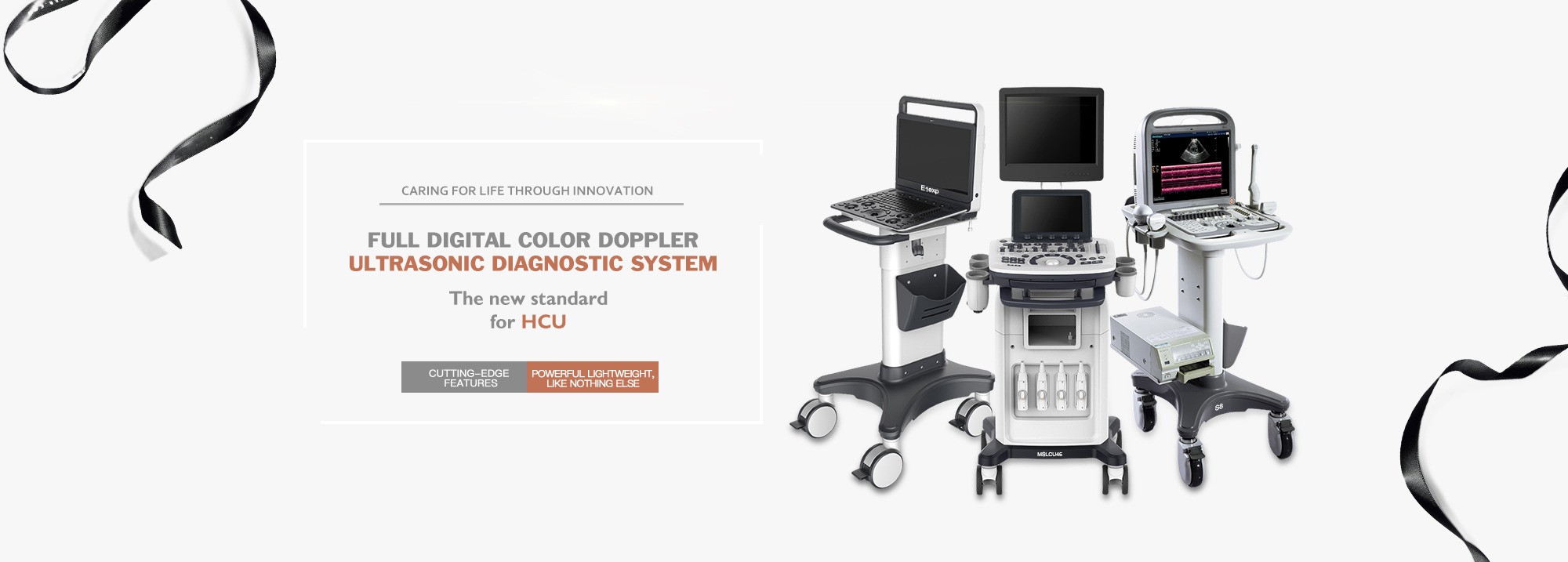
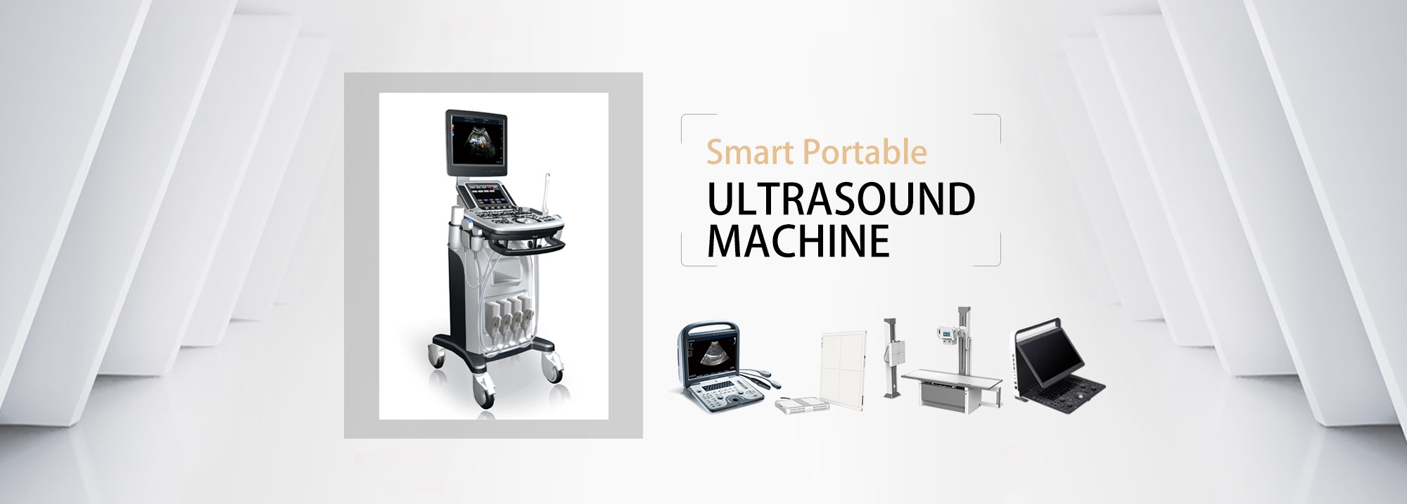
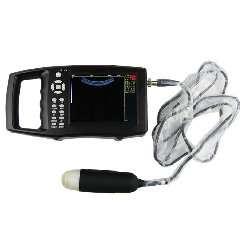
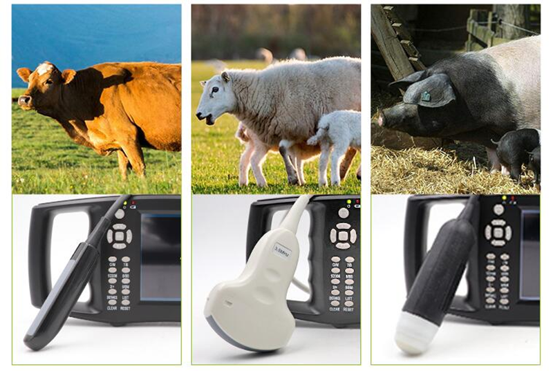
Ultrasound equipment is commonly used in pig farms, especially for breeding farms, which can be used to measure pregnancy, backfat, eye muscle, and some equipment to repel birds and animals is also used in ultrasound. You may often use ultrasound equipment, but you may not know some of the relevant knowledge, this article is a simple review of ultrasound technology used in pig farms.
Ultrasound
Ultrasound is a high-frequency sound wave, the range of the human ear to feel the sound wave is 20Hz to 20KHz, more than 20KHz (vibration 20 thousand times a second) sound wave is beyond the range of human hearing can feel, so it is called ultrasound.
The sound wave used by general ultrasound equipment is much higher than 20KHz, such as the frequency of the general electronic convex array ultrasound pregnancy scanner is 3.5-5MHz.
The reason why ultrasound will be used to detect equipment is mainly because of its good directivity, strong reflection, and certain penetration ability. The essence of ultrasound equipment is a transducer, which converts electrical signals into ultrasound waves to be emitted, and the ultrasound waves reflected back are received by the transducer, which are converted into electrical signals, and the electrical signals are further processed to form images or sounds.
A ultrasound
A-Ultrasound equipment is commonly used in pig farms, especially for breeding farms, which can be used to measure pregnancy, backfat, eye muscle, and some equipment to repel birds and animals is also used in ultrasound. You may often use ultrasound equipment, but you may not know some of the relevant knowledge, this article is a simple review of ultrasound technology used in pig farms.
Ultrasound
Ultrasound is a high-frequency sound wave, the range of the human ear to feel the sound wave is 20Hz to 20KHz, more than 20KHz (vibration 20 thousand times a second) sound wave is beyond the range of human hearing can feel, so it is called ultrasound.
The sound wave used by general ultrasound equipment is much higher than 20KHz, such as the frequency of the general electronic convex array ultrasound pregnancy scanner is 3.5-5MHz.
The reason why ultrasound will be used to detect equipment is mainly because of its good directivity, strong reflection, and certain penetration ability. The essence of ultrasound equipment is a transducer, which converts electrical signals into ultrasound waves to be emitted, and the ultrasound waves reflected back are received by the transducer, which are converted into electrical signals, and the electrical signals are further processed to form images or sounds.
A ultrasound
Since the motor rotation frequency has an upper limit, the B-ultrasound of the mechanical probe will have a limit in clarity. In order to obtain higher resolution, electronic probes have been developed. Instead of using a mechanically driven transducer to swing, the electronic probe places a number of "A-ultrasound" (flashlights) in a convex shape, each of which is called an array element. The current controlled by the chip excises each array in turn, thereby obtaining a faster signal sending and receiving frequency than a mechanical probe.
But sometimes you will find that some electronic convex array probes have worse imaging quality than good mechanical probes, which involves the number of arrays, that is, how many arrays are used together, 16? 32 of them? 64 of them? 128? The more elements, the clearer the image. Of course, the concept of channel number is also involved.
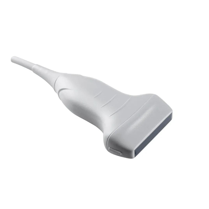
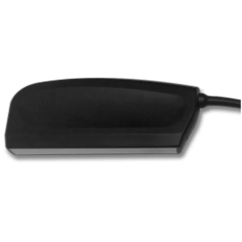
Further, you will find that whether the mechanical probe or the electronic convex array probe, the image is a sector. The near image is small, and the far image will be stretched. After the interference of transmitting and receiving signals between the array elements is technically overcome, the array elements can be lined up into a straight line, and the electronic linear array probe is formed. The image of the electronic array probe is a small square, just like the photo. Therefore, when using linear array probes to measure backfat, the three-layer lamellar structure of backfat can be perfectly presented.
By making the linear array probe a little larger, you get the eye muscle probe. It can illuminate the entire eye muscle, and of course, due to the relatively high price of the equipment, it is often only used in breeding.
Are there C-ultrasounds and D-ultrasounds?
No C-ultrasounds, but there's D-ultrasounds. D ultrasound is doppler ultrasound, is the application of doppler principle of ultrasound. We know that sound has a doppler effect, which is when a train passes in front of you, the sound goes faster and then slower. Using doppler's principle, he can let you know whether something is moving towards you or away from you. For example, when using ultrasound to measure blood flow, two colors can be used to mark the flow of blood, and the color depth is used to indicate the blood flow. This is called color ultrasound.
Color ultrasound and false color
There are many people who sell B-ultrasound will advertise that their products are color ultrasound. Clearly not the color ultrasound (D-ultrasound) we talked about in the previous paragraph. This can only be called fake color. The principle is like a black and white TV with a layer of color film. Each point on the B-ultrasound represents the intensity of the reflected signal at that distance, expressed in gray scale, so which color is essentially the same.
A-ultrasound can be compared to one-dimensional code (bar code); B-ultrasound can be compared to two-dimensional code, with false color B-ultrasound is painted two-dimensional code; D-ultrasound can be compared to three-dimensional code.
Post time: Jan-08-2024

