Some people say that the liver is the introduction to ultrasound, so the thyroid should also be the introduction to superficial ultrasound.
Ultrasound is no longer a simple picture and talk, ultrasound department is not a simple "auxiliary department" or "medical technology department", we are not only the clinical eyes, but also the active diagnosis after listening to the patient's main complaint, sometimes also often in the doctor's order to check some additional parts for patients free of charge, mainly to determine the diagnosis in our hearts, in order to clearly diagnose the disease, The normal condition of a certain organ is what we must master. Although the thyroid organ is small, there are many diseases. In order to make a true diagnosis, the ultrasound must not only master the normal anatomy and normal ultrasonic manifestations, but also master the etiology and main characteristics of differential diagnosis. Today we will first learn about normal thyroid and ultrasound manifestations:
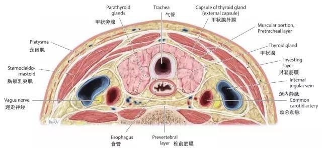
1. Anatomy of the thyroid gland
The thyroid is the largest endocrine gland in the human body, and its main function is to synthesize, store and secrete thyroxine.
The thyroid gland is located below the thyroid cartilage, on either side of the trachea, and consists of a central isthmus and two lateral lobes.
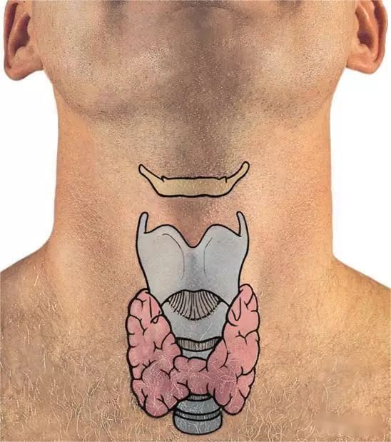
Thyroid body surface projection
Thyroid blood supply is very rich, mainly by the superior thyroid artery and inferior thyroid artery supply on both sides.
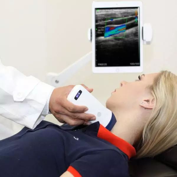
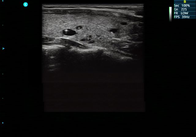
Ultrasound image of normal thyroid gland
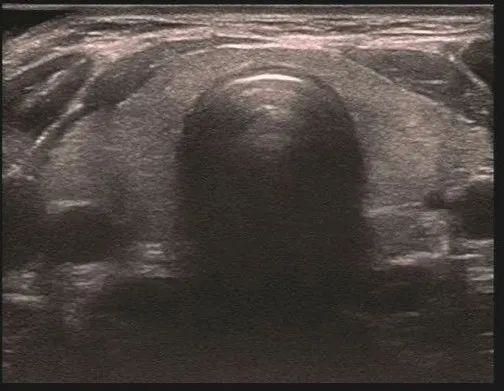
Cervical transthyroid section
2. Body position and scanning method
① The patient is in a supine position and lifts the lower jaw to fully extend the neck.
② When observing the lateral leaf, the face is facing the opposite side, which is more convenient for scanning.
③ The basic scanning methods of thyroid gland include longitudinal scan and transverse scan. First, the whole thyroid is examined in transverse section. After understanding the whole gland, the longitudinal section is examined.
3. Ultrasound findings of normal thyroid gland
Ultrasonically, the thyroid gland was in the shape of a butterfly or horseshoe, and the two sides of the lobe were basically symmetrical and connected to the central elongated isthmus. The trachea is located in the rear center of the isthmus, showing an arc of strong light with echo. The internal echo is medium, evenly distributed, with a thin dense light spot, and the peripheral muscle group is low echo.
Normal thyroid value: anterior and posterior diameter: 1.5-2cm, left and right diameter: 2-2.5cm, upper and lower diameter: 4-6cm; The diameter (thickness) of isthmus is 0.2-0.4cm
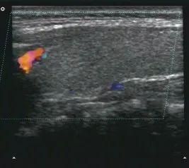
CDFI: Visible linear or speckled blood flow display, peak systolic velocity of arterial spectrum 20-40cm/s
Post time: Sep-21-2023


