Ultrasound examination is one of the most common examination methods and is an "essential" item for everyone's physical examination. So what is ultrasonic examination... Today we will take a closer look at ultrasonic examination to answer common questions.
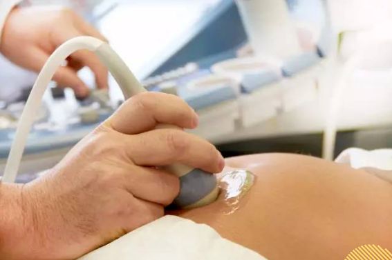
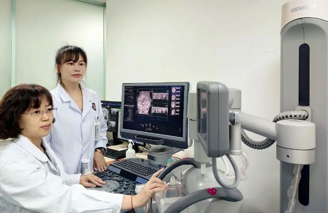
Ultrasound medicine, as an imaging medicine with rapid technological development in recent years, plays an irreplaceable role in the diagnosis and determination of treatment plans in clinical departments. Ultrasound guidance, interventional diagnosis and treatment play an important role in meeting the clinical minimally invasive needs. Recently, the Diagnostic Ultrasound Department has been equipped with a new ultrasound equipment with a laparoscopic probe. The following is an introduction to the imaging information and interventional treatments that the Diagnostic Ultrasound Department of our hospital can provide for clinical use in promoting the high-quality development of hospital disciplines.
1. Accurate diagnosis
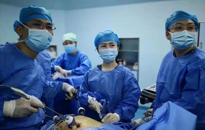
Laparoscopic probe shape and surgery The laparoscopic device is similar, except that a high-frequency ultrasound probe with adjustable direction is installed at the tip. It can directly enter the abdominal cavity through a hole in the abdominal wall to reach the surface of the organs for scanning, which is beneficial to accurately determine the location and surrounding proximity of the tumor during laparoscopic surgery. important vascular relationships.

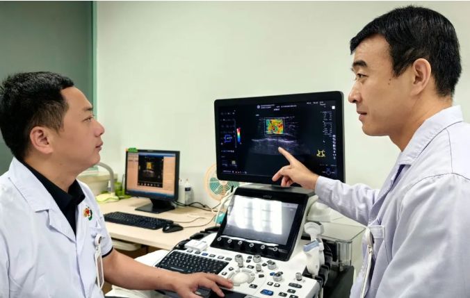
Contrast-enhanced ultrasound can determine the benign and malignant nature of space-occupying lesions in various parts. The intravenous injection of ultrasound contrast agent can improve the difference between the space-occupying lesions and the background echo. Compared with contrast-enhanced CT and MRI, contrast agents are metabolized through lung respiration and have negative effects on the liver and kidneys. It is also suitable for patients with functional impairment. Ultrasound elastography uses shear wave quantitative measurement to judge the hardness of superficial breast, thyroid and other tissue occupied areas, and then evaluate the benign and malignant properties of the occupied areas. Ultrasound elastography can also detect diffuse lesions such as liver cirrhosis and Hashimoto's thyroid. Yan et al. conducted quantitative analysis. Parametric imaging analyzes the blood perfusion inside the tumor to obtain fine perfusion time parametric imaging pictures that cannot be distinguished by the naked eye.
For example:
Ultrasound-guided intrahepatic bile duct drainage
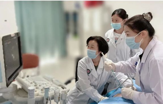
② Intraoperative laparoscopic ultrasound assists hepatobiliary surgery in precise liver resection Ultrasound elastography to evaluate musculoskeletal neuropathy
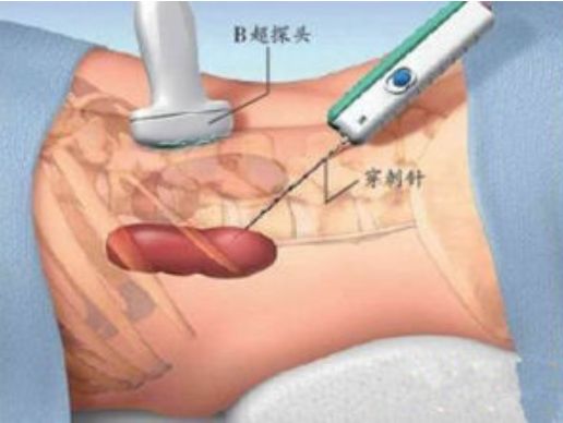
For puncture biopsy of tumors in various parts under ultrasound guidance, the position of the puncture gun needle tip can be observed in real time under ultrasound guidance, and the sampling angle can be adjusted at any time to obtain satisfactory specimens. The images generated by the Automatic Breast Volume Imaging System (ABVS) are three-dimensionally reconstructed, and the scanning process is standardized, which can more clearly display the lesions in the breast ducts. For smaller ducts, the coronal section can be observed, improving the diagnostic accuracy. Higher than ordinary two-dimensional breast ultrasound
For example:
Ultrasound-guided kidney biopsy
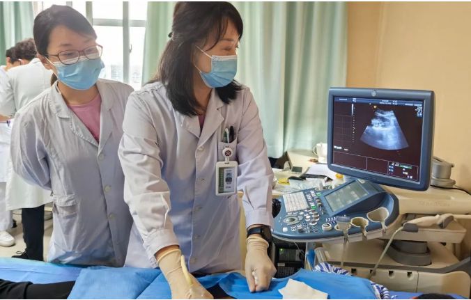
② Breast Automatic Volume Imaging System (ABVS) to detect lesions in breast ducts
2. Precision treatment
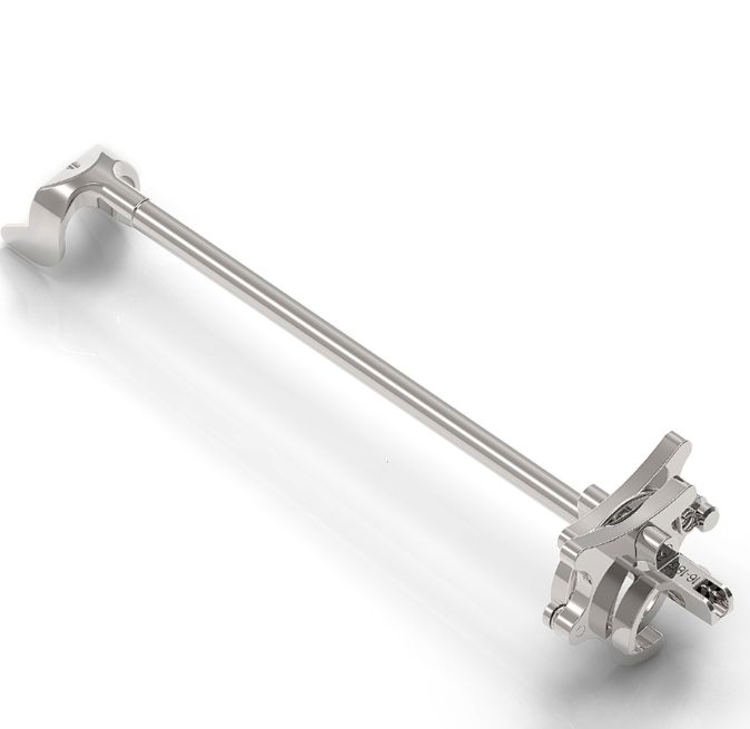
Ultrasound-guided ablation treatment of tumors is a minimally invasive and precise method to eliminate tumors. It causes minimal damage to the patient and is as effective as surgical resection. Comparable. Ultrasound-guided catheter drainage in various parts, especially the intrahepatic bile ducts, can monitor the positions of puncture needles, guide wires and drainage tubes in real time throughout the entire process without blind spots, and effectively and accurately insert drainage catheters to prolong the lives of patients with end-stage cholangiocarcinoma and improve their health. Quality of Life. Ultrasound-guided catheter drainage in the surgical area, chest, abdominal cavity, pericardium, etc. can minimally invasively relieve the pressure of fluid accumulation in various parts. Puncture biopsy guided by contrast-enhanced ultrasound can accurately take samples from the hyperperfused (active) area of the tumor to obtain satisfactory pathological results. With the widespread development of clinical endovascular interventional diagnosis and treatment, the occurrence of pseudoaneurysms is inevitable. Ultrasound-guided pseudoaneurysm sealing treatment can observe the effect of injecting thrombin in real time, so as to achieve satisfactory sealing with the smallest drug dose. effect and avoid complications to the greatest extent
Post time: Nov-03-2023

