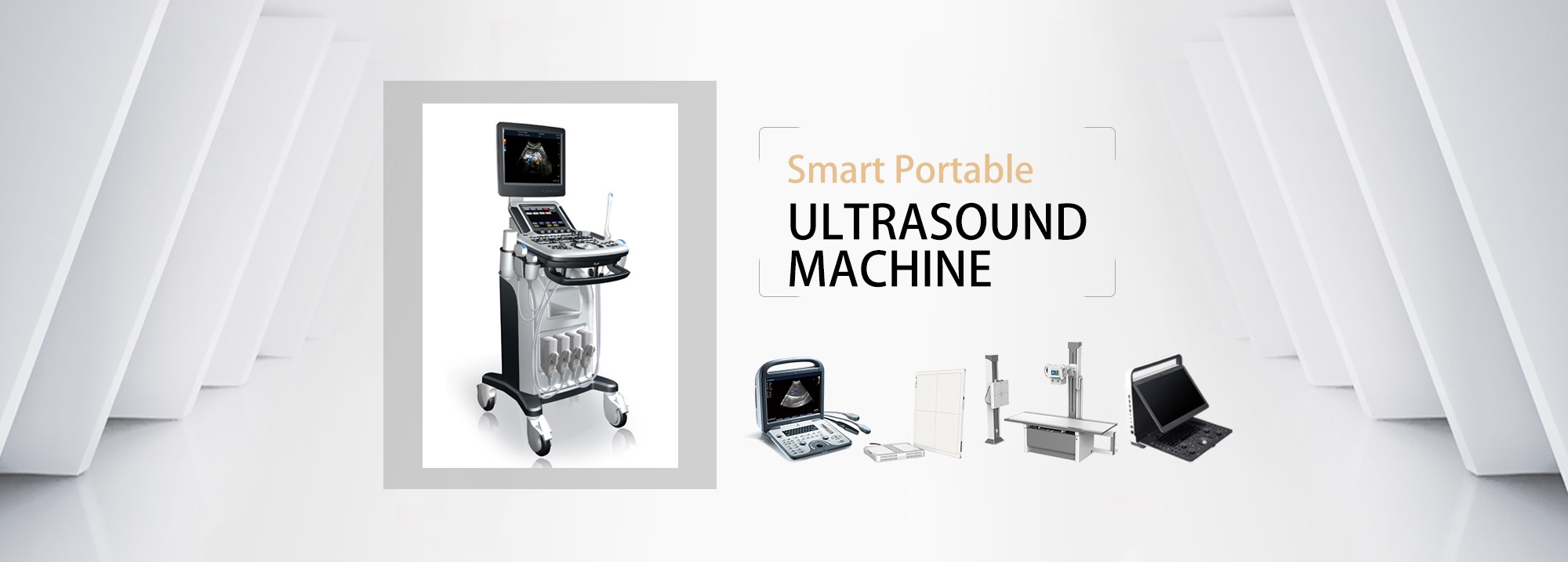
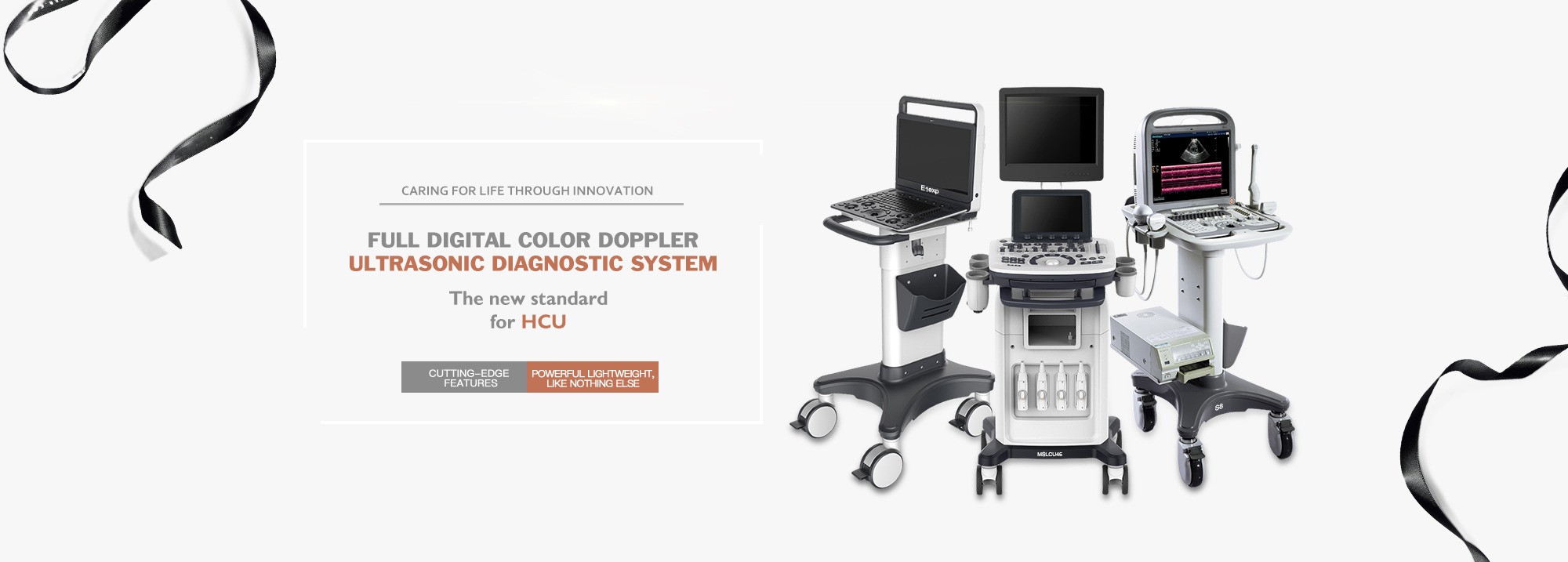
Ultrasonic imaging diagnosis technology has been developing for more than half a century in China. With the continuous improvement of electronic information technology and computer imaging technology, ultrasonic diagnostic equipment has also been revolutionary development for many times, from analog signal/black and white ultrasound/harmonic contrast/artificial recognition, to digital signal/color ultrasound/elastic imaging/artificial intelligence. New functions and application levels continue to expand, and ultrasonic imaging diagnostic equipment continues to innovate and break through, prompting the medical industry to have a huge demand for it.
01. Basic classification of common ultrasonic imaging diagnostic equipment
Ultrasonic imaging diagnostic equipment is a kind of clinical diagnostic equipment developed according to the principle of ultrasound. Compared with large medical equipment such as CT and MRI, its inspection price is relatively low, and it has the advantages of non-invasive and real-time. Therefore, the clinical application is more and more extensive. At present, ultrasound examination is roughly divided into A-type ultrasound (one-dimensional ultrasound), B-type ultrasound (two-dimensional ultrasound), three-dimensional ultrasound and four-dimensional ultrasound.
Usually referred to as B-ultrasound, it actually refers to black and white two-dimensional B-ultrasound, the collected image is a black and white two-dimensional plane, and color ultrasound is the collected blood signal, after the computer color coding on the two-dimensional image in real time superposition, that is, the formation of color Doppler ultrasound blood image.
Three-dimensional ultrasonic diagnosis is based on color Doppler ultrasonic diagnosis instrument, data acquisition device is configured, and image reconstruction is carried out through three-dimensional software, so as to form a medical device that can display three-dimensional imaging function, so that human organs can be displayed more stereoscopic and lesions can be found more intuitively. The four-dimensional color ultrasound is based on the three-dimensional color ultrasound plus the time vector of the fourth dimension (inter-dimensional parameter).
02. Ultrasonic probe types and applications
In the process of ultrasonic image diagnosis, ultrasonic probe is an important part of ultrasonic diagnosis equipment, and it is a device that transmits and receives ultrasonic waves in the process of ultrasonic detection and diagnosis. The performance of the probe directly affects the characteristics of ultrasonic and ultrasonic detection performance, so the probe is particularly important in ultrasonic image diagnosis.
Some conventional probes in ultrasonic probes mainly include: single crystal convex array probe, phased array probe, linear array probe, volume probe, cavity probe.
1, single crystal convex array probe
The ultrasonic image is the product of the close combination of the probe and the system platform, so on the same machine, the software and hardware need to meet the requirements of the single crystal probe.
The single crystal convex array probe adopts the single crystal probe material, the probe surface is convex, the contact surface is small, the imaging field is fan-shaped, and it is widely used in the abdomen, obstetrics, lungs and other relative parts of the deeper organs.
Liver cancer examination
2, phased array probe
The probe surface is flat, the contact surface is small, the near field field is minimal, the far field field is large, and the imaging field is fan-shaped, which is suitable for the heart.
Cardiac probes are usually divided into three categories according to the application population: adults, children, and newborns: (1) adults have the deepest heart position and slow beating speed; (2) The position of the newborn heart is shallow and the beating speed is the fastest; (3) The condition of children's hearts is between that of newborns and adults.
Cardiac examination
3, linear array probe
The probe surface is flat, the contact surface is large, the imaging field is rectangular, the imaging resolution is high, the penetration is relatively low, and it is suitable for superficial examination of blood vessels, small organs, musculoskeletal and so on.
Thyroid examination
4, volume probe
On the basis of two-dimensional image, the volume probe will continuously collect the spatial distribution position, through the computer reconstruction algorithm, so as to obtain the complete spatial shape. Suitable for: fetal face, spine and limbs.
Fetal examination
5, cavity probe
The intracavitary probe has the characteristics of high frequency and high image resolution, and does not need to fill the bladder. The probe is close to the examined site, so that the pelvic organ is in the near field area of the sound beam, and the image is clearer.
Examination of endovascular organs
Post time: Aug-23-2023


