To clarify these two issues, we must first know the definition of ultrasound, which is a type of medical imaging. See medical ultrasound for their classification:
| Medical Imaging | |||
|
|
|
Medical | ● Pneumoencephalography imaging
● Upper gastrointestinal series/Small-bowel follow-through/Colon barium enema ● Cholangiography/Cholecystography ● Mammography ● Angiography |
| Commercial | Radiographic testing | ||
|
CT |
Technology | ● General operation of CT● Quantitative CT● High resolution CT | |
|
Target |
● coronary artery● Calcium scan● CT angiography
● Computed Tomography Angiography ● Cranial computed tomography |
||
| Other | ● Fluoroscopy● X-ray motion analysis | ||
|
Magnetic Resonance |
● MRI of the brain● MR neurography● Cardiovascular Magnetic Resonance Imaging/Cardiac MRI perfusion
● Functional Magnetic Resonance Imaging ● Diffusion MRI |
||
|
|
● Echocardiography● Doppler ultrasound● Doppler echocardiography
● Gynecology ● Abdomen ● Contrast Ultrasound Imaging |
||
|
nuclear medicine |
2D / scintigraphy | ||
| 3D/ECT |
|
||
| Masako contrast(PET)1. Brain PET 2.Cardiac PET 3.PET mammography 4.PET-CT 5.PET-MRI |
|||
|
Optical/Laser |
|
||
|
Thermal Imager |
|||
1.Ultrasound definition:
Ultrasound is medical sonography (ultrasound, diagnostic sonography), an ultrasound-based medical imaging diagnostic technique that enables visualization of soft tissues such as muscles and internal organs, including their size, structure, and pathological lesions. Obstetric ultrasonography is widely used for prenatal diagnosis during pregnancy.
Scientists refer to the number of vibrations per second as the frequency of sound, and it is measured in hertz (Hz). The frequency of sound waves that our human ear can hear is 20Hz-20000Hz. Therefore, we call sound waves with frequencies above 20,000 Hz “ultrasonic”. Ultrasound frequencies typically used for medical diagnosis are 1 MHz-30 MHz.
While the term “ultrasound” is used in physics to refer to all frequencies above the upper limit of the human hearing threshold (20,000 Hz, 20 kHz), in medical imaging it usually refers to sound waves with a frequency band more than a hundred times higher.
2.Principle of Ultrasound: Ultrasound propagates in human tissue and will produce reflection, transmission, refraction and scattering. In addition, the relative movement of the ultrasonic transmitting device and the human body will also produce the ultrasonic Doppler phenomenon. Thus, three working principles of ultrasonic diagnostic instruments are formed, namely pulse echo principle, ultrasonic Doppler principle and transmission principle.
3. Ultrasound classification: There are four types (modes) of medical ultrasonography: A-mode (Amplitude-mode), B-mode (Brightness-mode), M-mode (Motion-mode), Doppler-mode (Doppler-mode)
A-A mode is the simplest type of ultrasound, a single sensor scans a line through the body and the echoes are plotted on the screen as a function of depth; therapeutic ultrasound for a specific tumor or stone is also A-mode, allowing precise localization of destructive wave energy .
B-B mode ultrasound, a linear array of transducers scans a plane across the body simultaneously, allowing a two-dimensional image to be seen on the screen.
M- M mode ultrasound (M stands for movement), rapid sequences of B-mode scans have images displayed sequentially on the screen as the boundary of the organ that produces the reflection moves relative to the probe, allowing the physician to see and measure the range of motion.
Doppler- mode uses the Doppler effect to measure and display blood flow and can be used to assess whether a structure (usually blood) is moving towards or away from the probe, and its relative velocity, which is especially useful in cardiovascular research. Ultrasounds used for diagnosis include black and white ultrasound and color ultrasound. Black and white ultrasound uses B-mode ultrasound imaging, so it is also called B-ultrasound; color ultrasound uses the Doppler principle.
4. Ultrasound industry brief: Mindray, Sonoscape , Chison, EDAN, WELLD, Emperor, SIUI, HAIYING in China’s domestic ultrasound; GE, PhilipsPhilips, Siemens among international brands ,Toshiba,Hitachi- Aloka,Esaote,SamSung- Medison,Sonosite
4. Scope of application: Ultrasonography is now widely used in medicine. It may be diagnostic, or it may be guiding during treatment (eg, biopsy or drainage of effusions). Typically a hand-held probe (often called a probe) is placed on the patient and moved to scan, and a water-based gel is applied to couple between the patient’s body and the probe.
5. The medical use of ultrasound has cardiology
● Endocrinology
● Gastroenterology (abdominal ultrasound)
● Gynecology; see Gynecologic Ultrasound
● Obstetrics; see Obstetric Ultrasound
● Ophthalmology; see Ultrasound A, Ultrasound B
● Urology
● Vascular
● CEUS
● Ophthalmology
● Pelvic ultrasound is the primary diagnostic tool for PCOS and can also be used to image the uterus, ovaries, and bladder. Ultrasound during pregnancy is used to check the development of the fetus. Men sometimes have a pelvic ultrasound to check the health of the bladder and prostate. Pelvic ultrasonography is performed in two ways: percutaneous and intracavitary. Intracavitary ultrasound can be transvaginal (women) or transrectal (men)
6. Advantages of Ultrasound
● Good visualization of muscle and soft tissue, especially useful for showing the interface between solid and liquid cavities;
● Real-time generation of images, inspection operators can dynamically select the most useful part to observe and record, which is conducive to rapid diagnosis;
● show the structure of the organs;
● There are currently no known long-term side effects, which generally do not cause patient discomfort;
● Equipment is widely distributed and relatively flexible;
● Small, portable scanners are available; examinations can be performed at the patient’s bedside;
● Inexpensive compared to other tests (eg CT imaging, bidirectional X-ray absorption imaging or MRI).
1.Let’s think about it, What is the future trend of ultrasound development:?
From the market point of view, with the rapid development of digital technology, Internet of Things technology and mobile intelligent terminal equipment, the intelligence and miniaturization of medical equipment will become the key development trend in the future. The ultra-small hand-held ultrasonic “inspector” of digital technology will eventually become the right-hand man of every doctor’s diagnosis and treatment, and even enter the home like electronic blood pressure monitors and electronic blood glucose meters, and become a routine inspection tool for people’s preventive health care. Therefore, it has a broad market space
2.How to find the best price ultrasound?
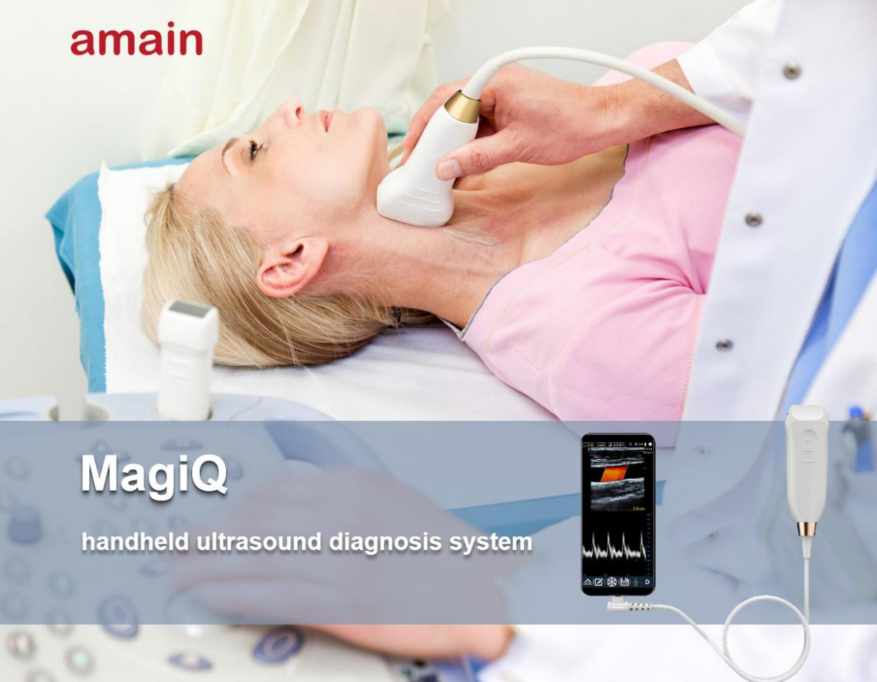
Amain handheld portable ultrasound—MagiQ series, bring Innovations for you, let you enjoy the intelligent life with Digital handheld probe-type ultrasound system
Fetures:
● Portable– The most portable devicesGets the traditional ultrasound machine smaller into a transducer. You can put it and your smart device with software into your pocket to anywhere
● Convenient– Easy to operateThe humanized ultrasound interface design, you can operate easily with your smart devices
● High resolution– Low power, consumption stable HD image
Image processing technology can offer you a high quality image.
● Mutipurpose–Wide applications, visible diagnostic apparatusit can be used in mutiple departments, such as: OB/GYN, Urology, Abdomen, Emergency, ICU, Small and shallow parts.
● Smart– Applicable to mutiple terminals Ultrasound app brings diagnostic capabilities to your compatible smartphone or handheld device.
Functions:
● Incredible image processing technology
Digital beaming forming, continuous dynamic focusing, and dynamic apodization. Healson portable ultrasound transducers and app include decades of expertise and innovation in ultrasound imaging to help you make fast, informed decisions.
● Report and image management
Freeze/real-time image storage, multiple image format storage such as png, jpeg and so on. Maximum 512 frames cineloop storage, USB disk storage, and DICOM 3.0. Plentiful report template, editing and saving report function.
● Humanized ultrasound interface design, easy to operateEnglish / Chinese, customized language available. Imaging Optimizing: Contrast adjust ment, brightness adjustment, Gamma adjustment, intelligent noise reduction, and abundant color package. Besides normal measurement, support professinal measurement including Abdominal, OB/GYN, Urology, Small Parts, and so on. Model:
1.Black and white ultrasound
● MUC5-2
● MUC5-2E
● MUEV9-4E
● MUL8-4E
● MUL8-4T
● MUL5-2ET
3. Color doppler ultrasound
MUL10-5S
MUL10-5
MUL5-2E
MUCL
MUL8-4T
MUL5-2ET
Post time: Jun-30-2022


