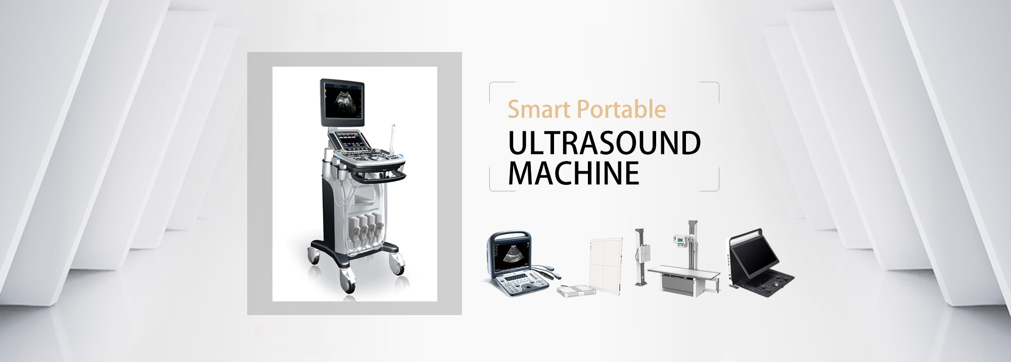
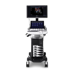
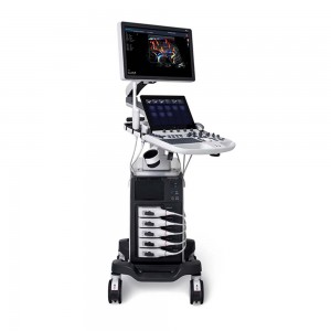
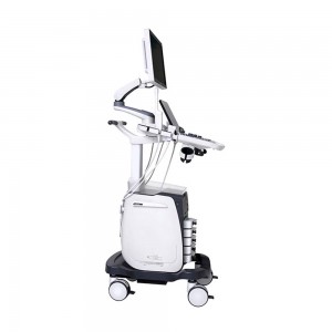
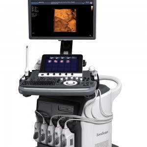
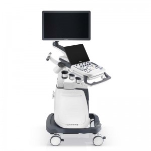
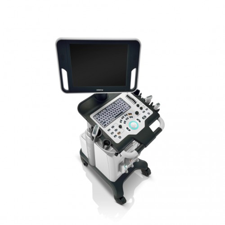
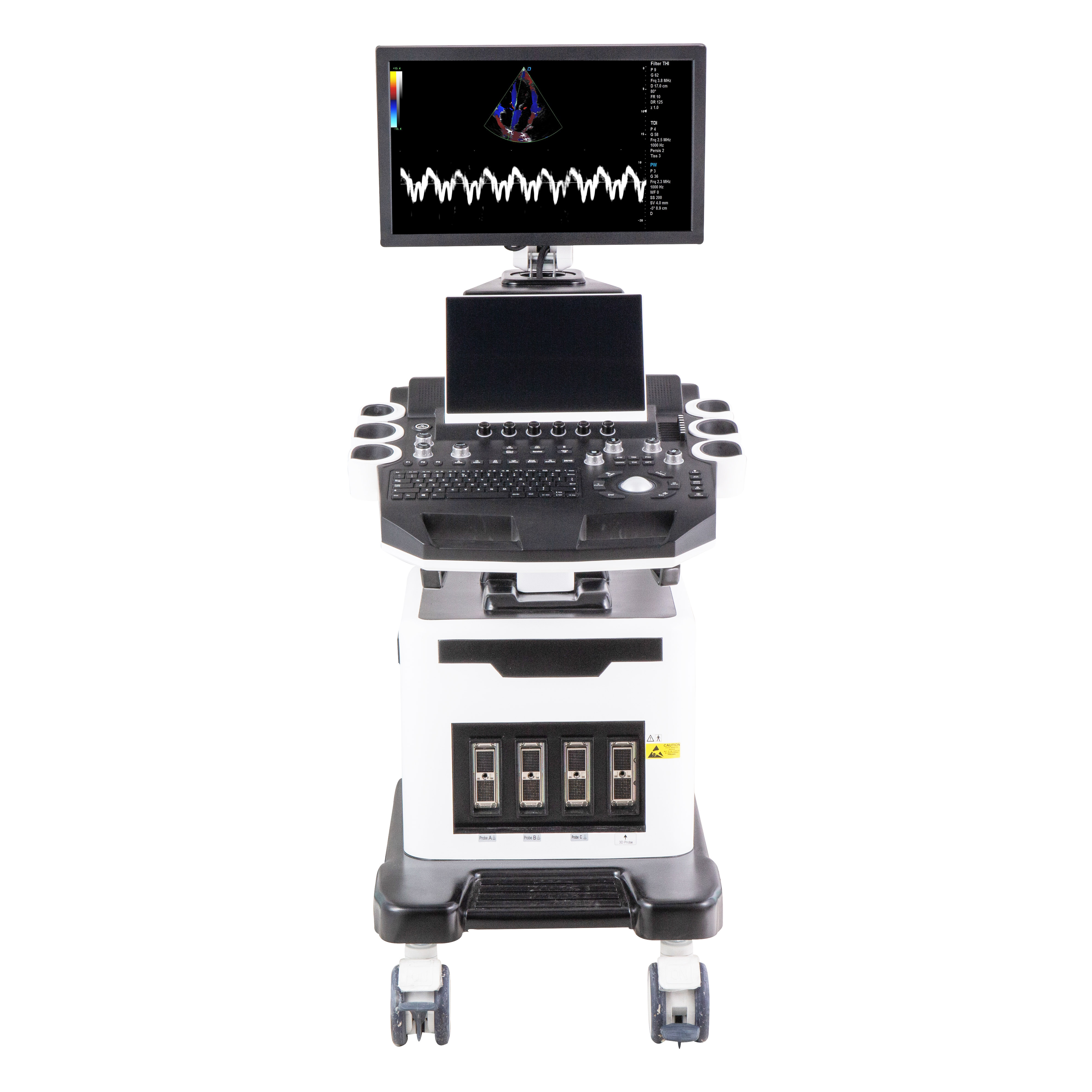
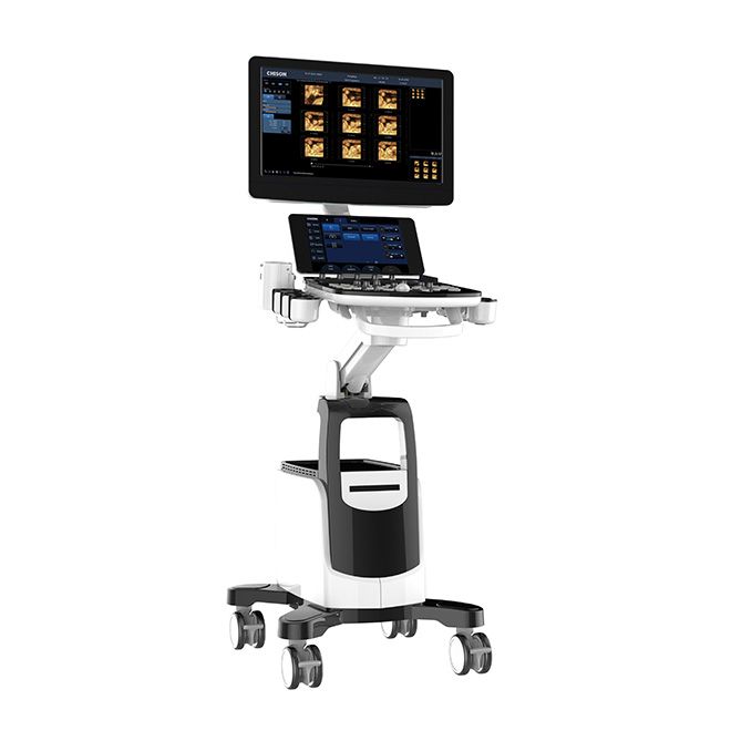
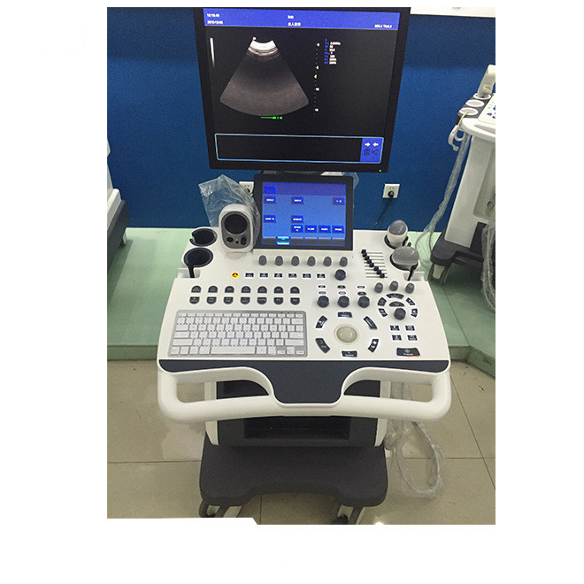
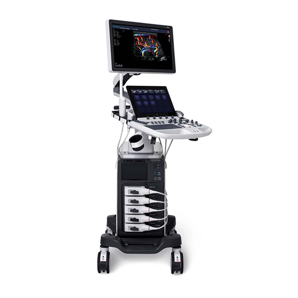
SonoScape P50 Elite integrates a number of new chips and ultra-integrated hardware modules to greatly improve the image frame rate. At the same time, CPU+GPU parallel processing technology is adopted to balance the performance of high-end system and small and flexible body. Its extreme processing speed, high-end application functions, rich probe collocation, will bring you unprecedented quality experience, so that ultrasound examination becomes more convenient and efficient.
Specification
|
21.5 inch high definition LED monitor
|
|
13.3 inch quick response touch screen
|
|
Height-adjustable and horizontal-rotatable control panel
|
|
Five Active Probe Ports
|
|
One Pencil Probe Port
|
|
External Gel Warmer (temperature adjustable)
|
|
Built-in ECG Module (Including Hardware and Software)
|
|
Built-in Wireless Adapter
|
|
2TB Hard Disk Drive, HDMI Output and USB 3.0 Ports
|


Product Features
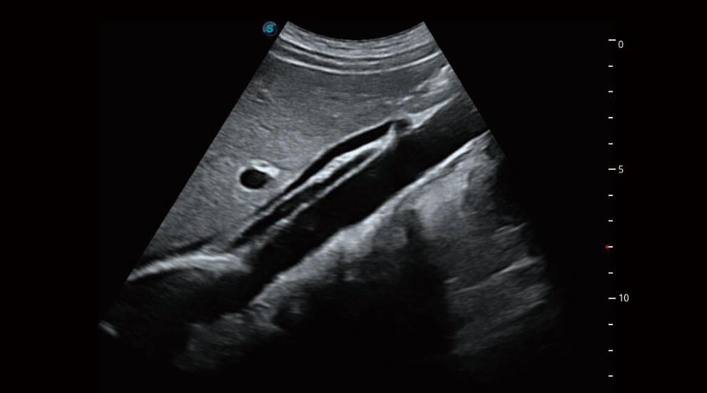
μScan+
Available for both B and 3D/4D modes, the new generation μScan+ provides you authentic presentation of details and lesion display through speckle reduction and enhanced border continuity.
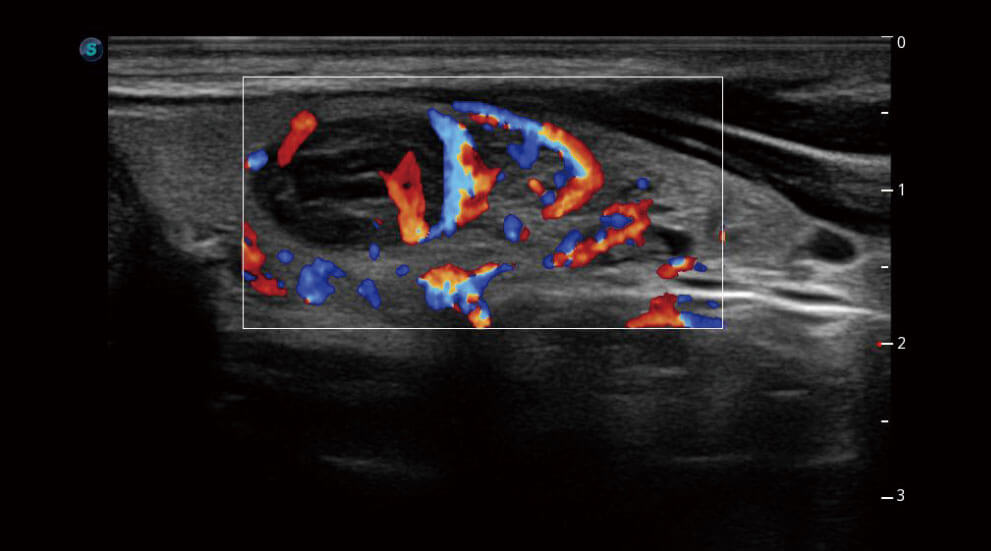
SR-Flow
Highly effective filter technology visualizes slow flows, enabling a vivid Doppler display with high sensitivity.
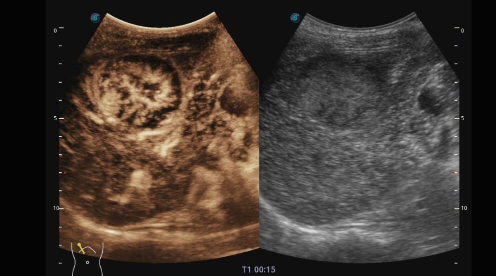
CEUS with MFI
Enhanced perfusion display traces small bubble populations, even in low-perfused and peripheral regions.
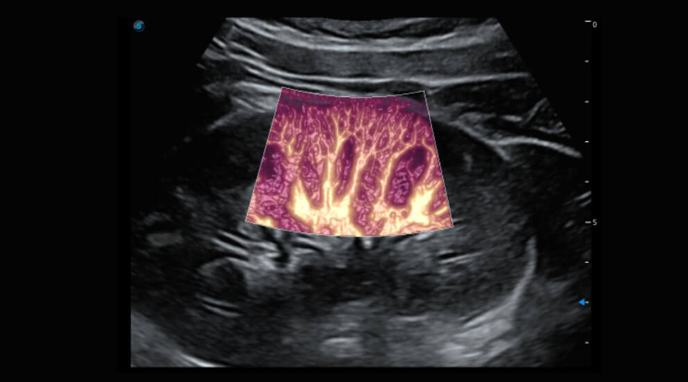
Bright Flow
3D-like color Doppler flow strengthens boundary definition of vessel walls, without the need of using volume transducer.
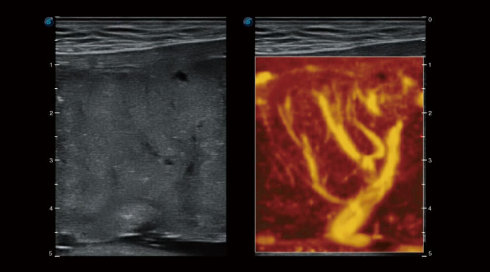
Micro F
Micro F provides an innovative method to expand the range of visible flow in ultrasound, especially for visualizing hemodynamic of tiny vessels.
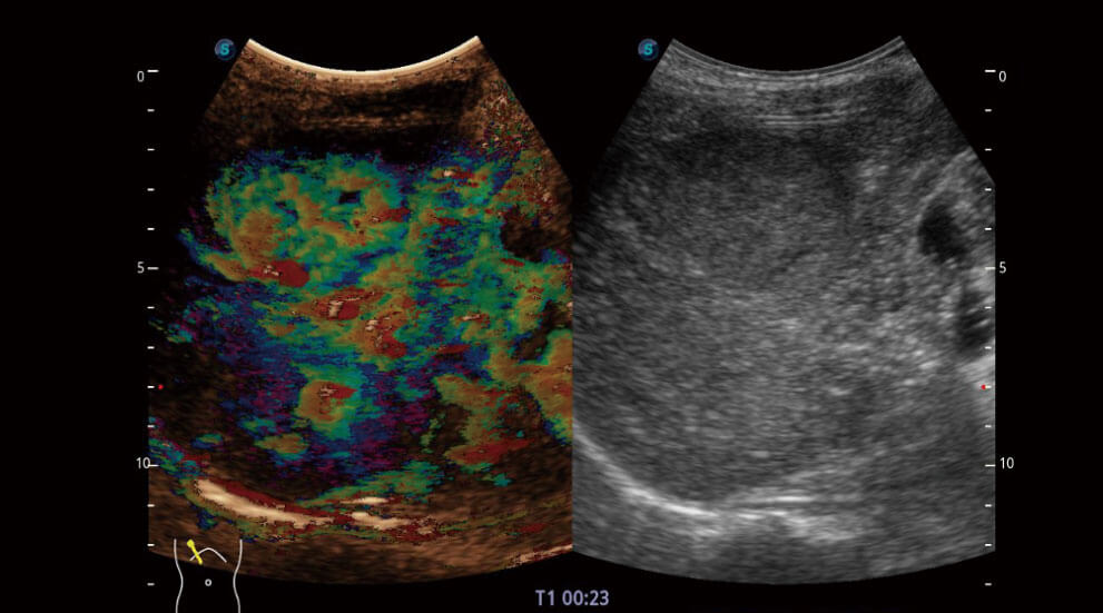
MFI-Time
To better differentiate tissues, color coded parametric view indicates the uptake time of contrast agents in different perfusion phases.
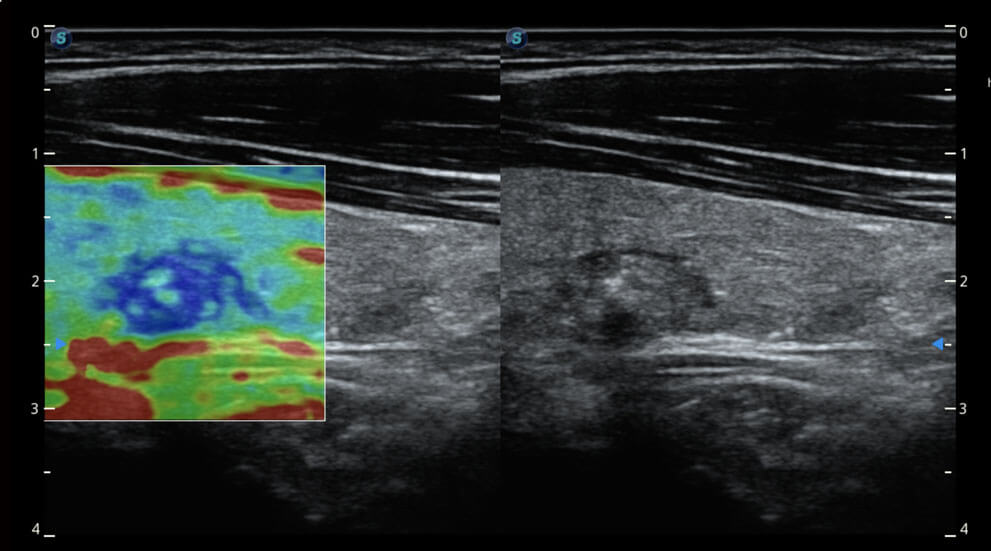
Strain Elastography
Real-time tissue stiffness assessment based on strain detects potential tissue abnormalities with an intuitive color map displayed. Semi-quantitative analysis of strain ratio indicates relative stiffness of the lesion.
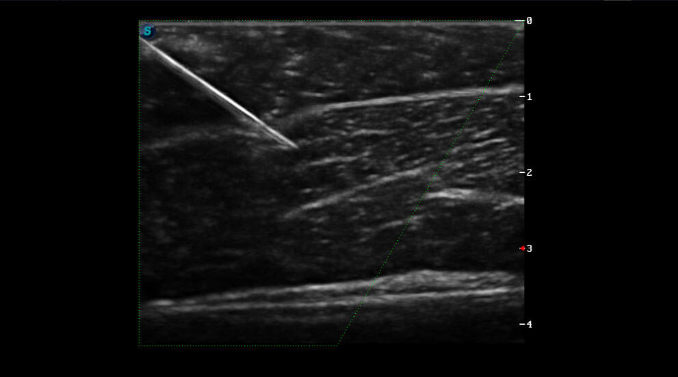
Vis-Needle
Improved accuracy and efficiency in diagnosis are possible with beam steering added to Vis-Needle, which provides enhanced visibility of the needle shaft and needle tip to assist with safe and accurate interventions such as nerve blocks.
ELITE in Cardiovascular
The caring for maternal and fetal well-being is underlying the conception of designing P50 ELITE. Outstanding 3D/4D imaging. Intelligent evaluation. Streamlined workflow. Those are the exact ways how P50 ELITE is transforming OB/GYN exams.
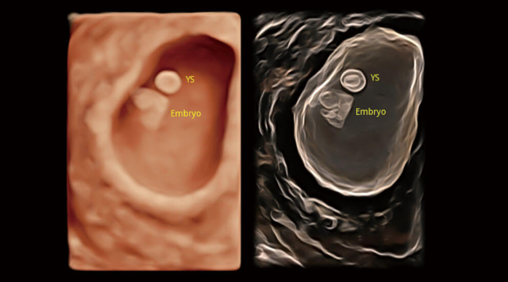
S-Live & S-Live Silhouette
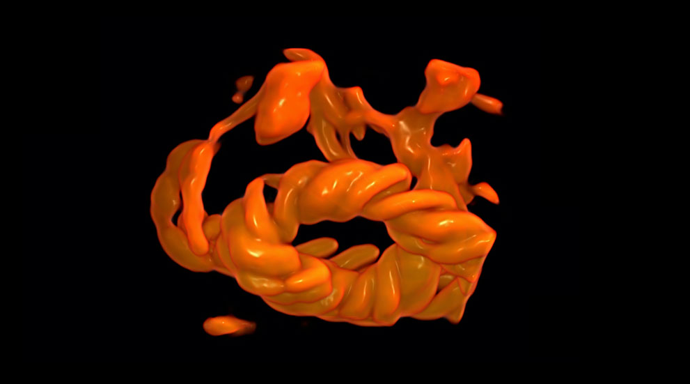
Color 3D
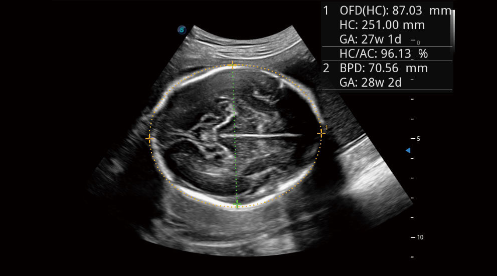
S-Fetus
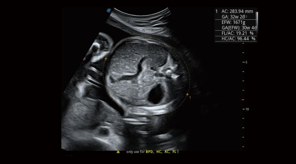
Auto OB
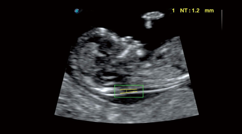
Auto NT
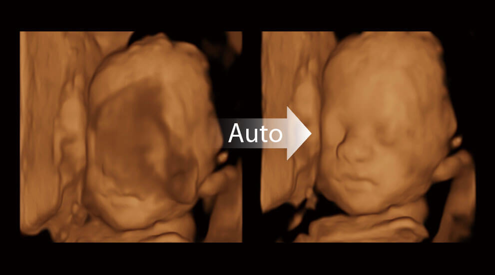
Auto Face
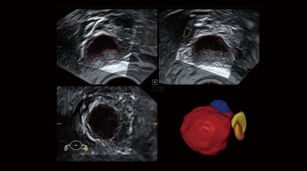
AVC Follicle
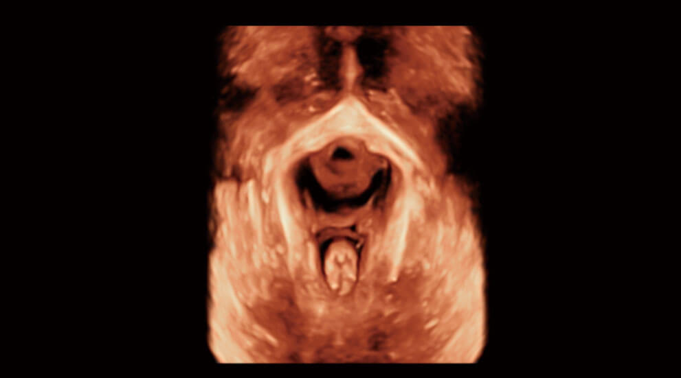
Pelvic Floor Imaging
ELITE in OB/GYN
P50 ELITE takes the following as its duty, visualize anatomy more confidently with enhanced 2D and color image quality; accelerate exams with automated expert tools; gain quantitative results with advanced capabilities for heart function assessment.
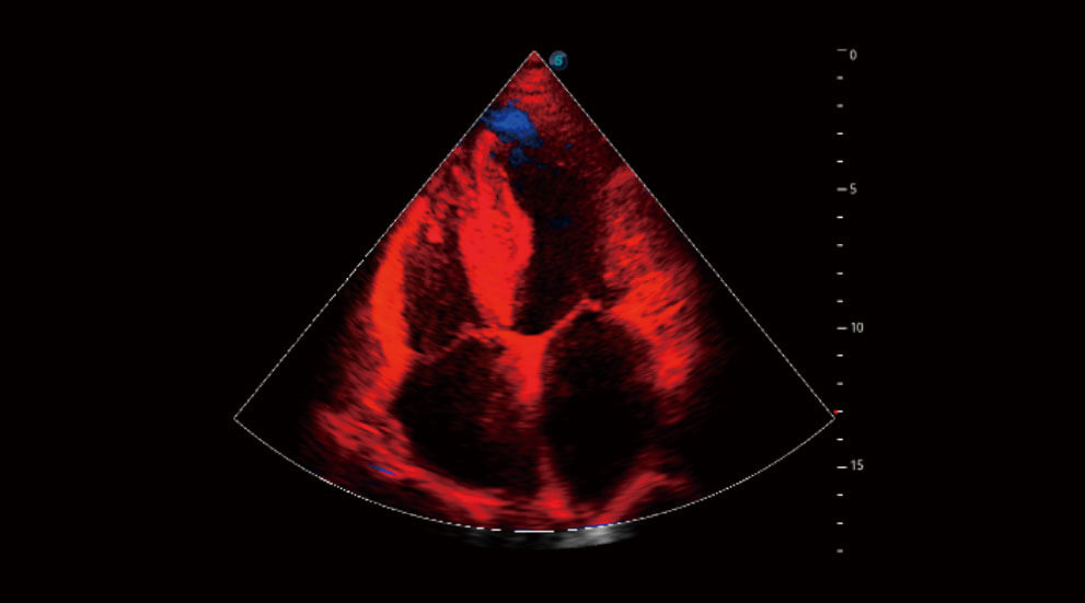
Tissue Doppler Imaging (TDI)
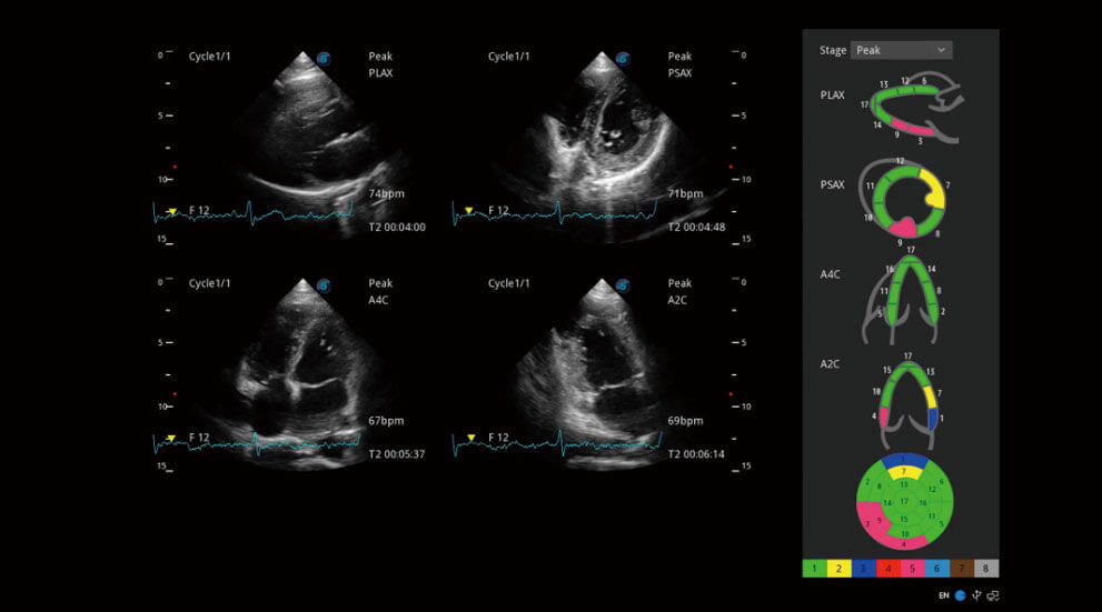
Stress Echo
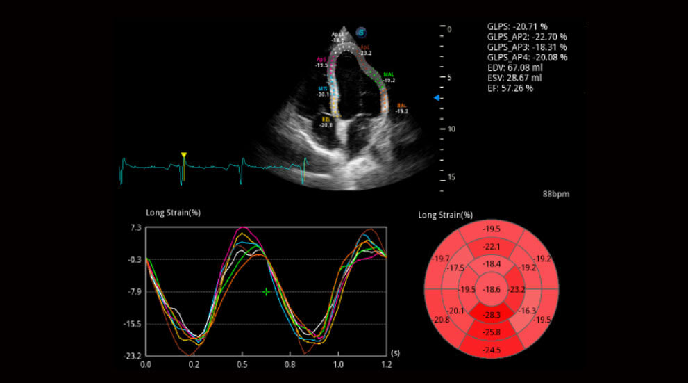
Myocardium Quantitative Analysis (MQA)
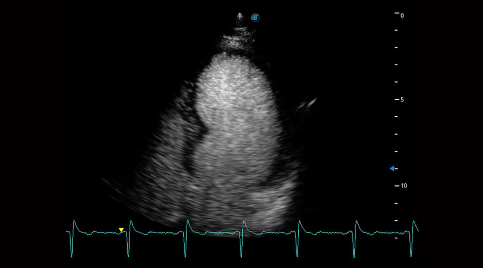
LVO
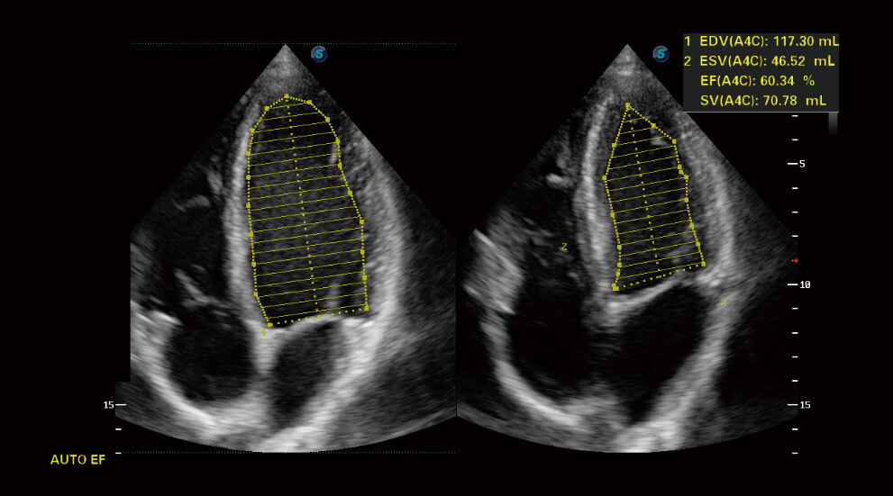
Auto EF
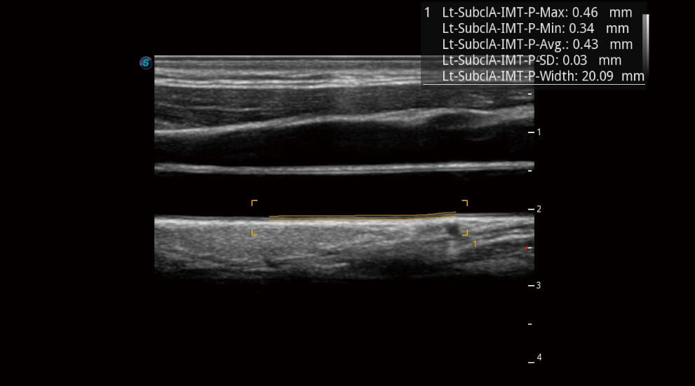
Auto IMT

