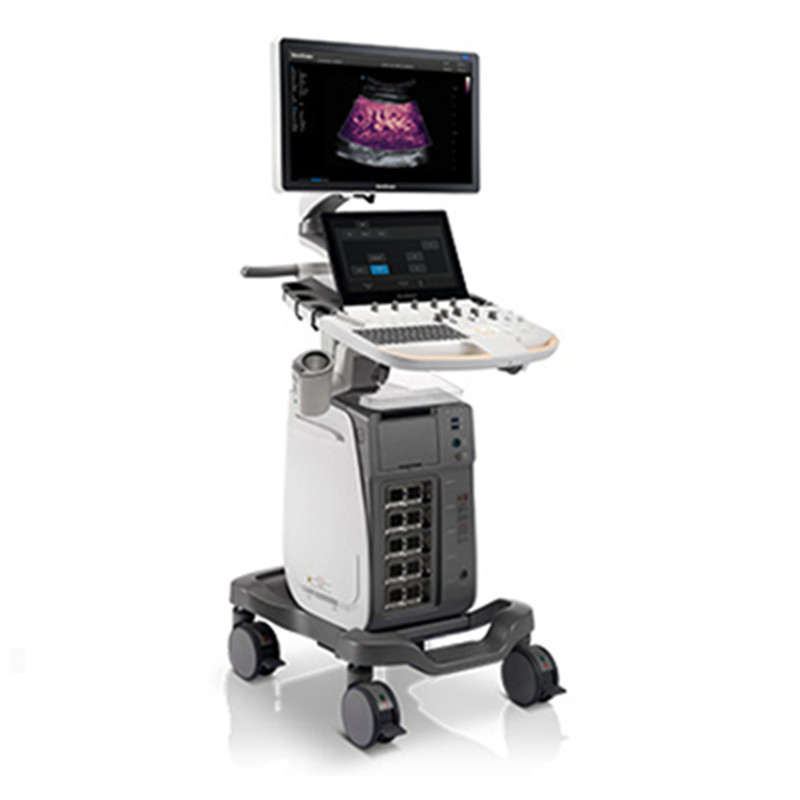
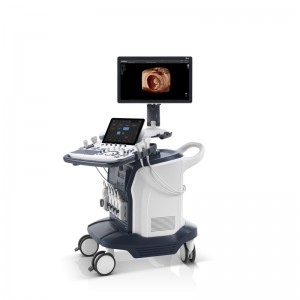
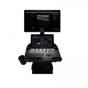
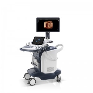
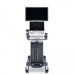
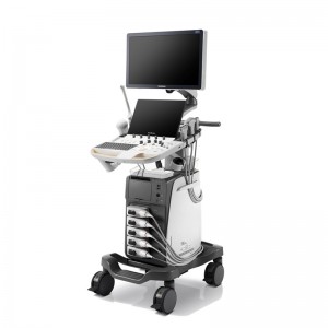
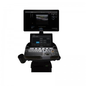
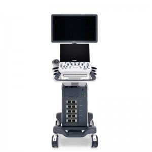
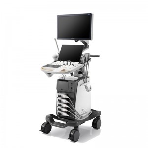
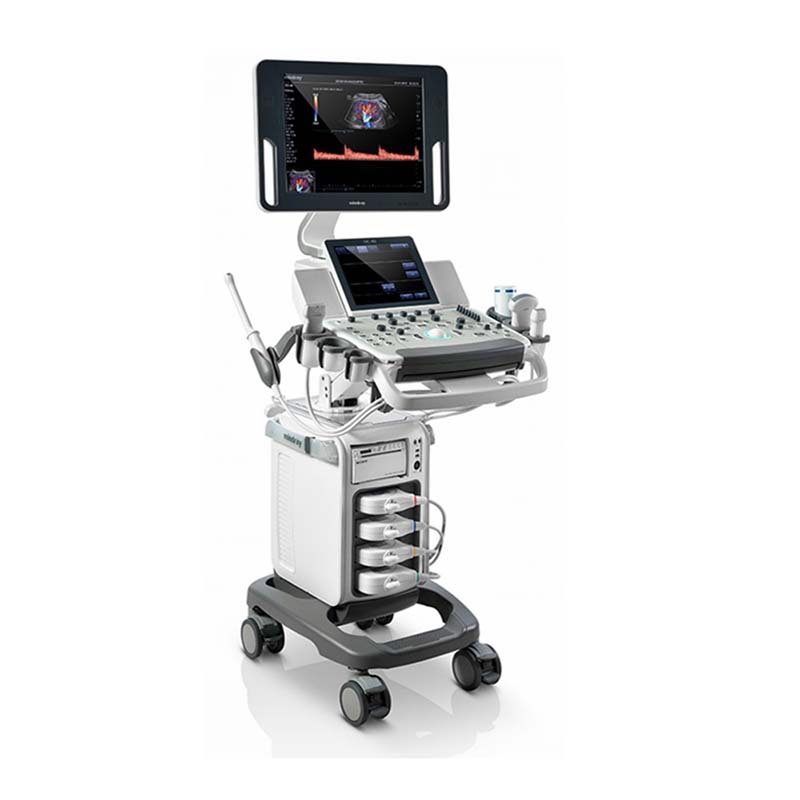
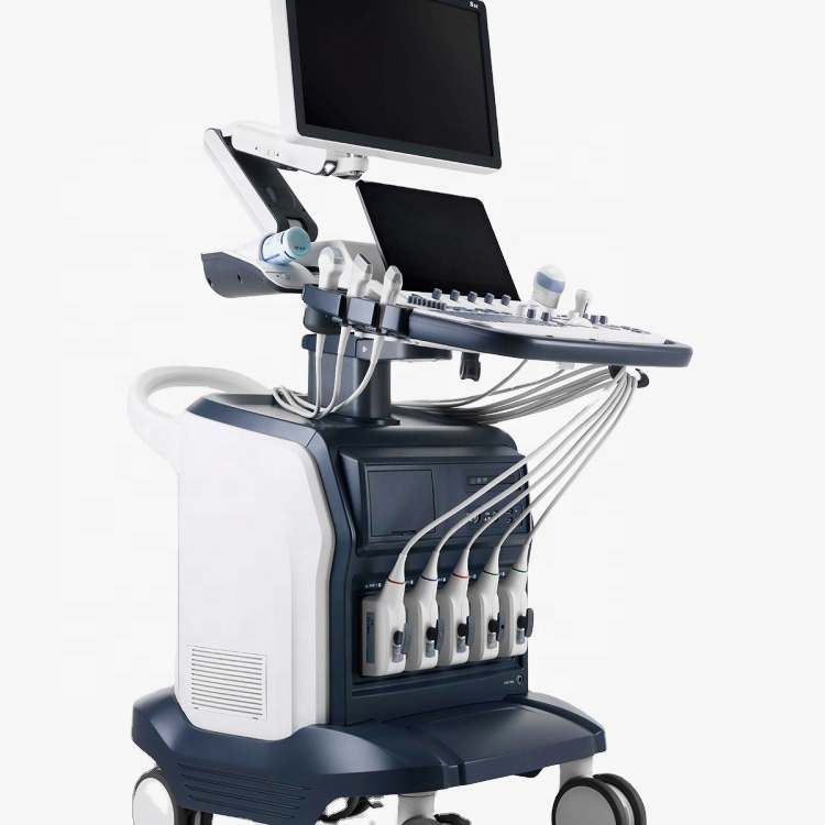
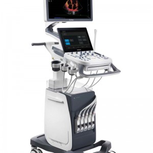
SonoScape P60 Cart-Based System Echocardiography Ultrasound Instruments With 7.5MHz Linear Transducer
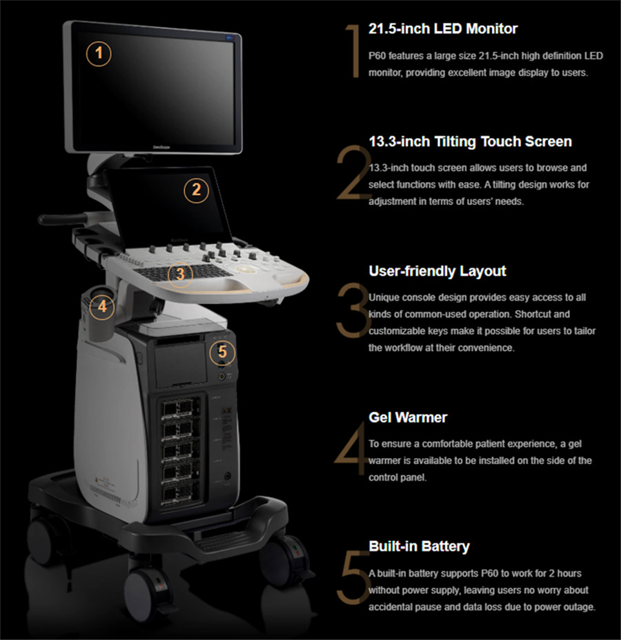
| Standard Configuration | P60 Main Unit 21.5" High Resolution Medical Monitor 13.3" High Resolution Touch Screen Height Adjustable and Rotatable Operation Panel Five Active Probe Ports One Pencil Probe Port Build-in ECG Module (Including Hardware and Software) External Gel Warmer (temperature adjustable) Built-in Wireless Adapter 1TB Hard Disk Drive, HDMI Output and USB 3.0 Ports |
| Imaging Mode | B (2B & 4B) Mode M Mode Anatomic M Mode Color M Mode Color Doppler Flow Imaging Power Doppler Imaging / Directional Power Doppler Imaging Tissues Doppler Imaging Pulse Wave Doppler Imaging Continuous Wave Doppler Imaging High Pulse Repeat Frequency Tissue Harmonic Imaging Pulse Inversion Harmonic Imaging Spatial Compound Imaging Tissue Specific Imaging Image Rotation μ-Scan: 2D Speckle Reduction Technology 3D μ-Scan: 3D Speckle Reduction Technology SR Flow (High Resolution Flow) Simultaneous Mode (Triplex) FreeHand 3D Imaging B Mode Panoramic Imaging / Color Panoramic Imaging Lateral Gain Compensation Trapezoid Imaging Widescan Imaging (Convex Extended Imaging) Biopsy Guide Vis-needle (Needle Visualization Enhancement) Auto Bladder Volume Measurement Zoom (Pan-Zoom / HD-Zoom / Scr-Zoom) TEI Index PW Auto Trace Auto IMT Auto EF Auto NT Auto OB: BPD / HC / AC / FL / HL S-Guide C-xlasto (Strain Elastography) Build-in User Mannual (Help) Sono-help (Scanning Tutorial) DICOM 3.0: Store / C-Store / Worklist / MPPS / Print / SR / Q&R |
| Applications | Basic Measurement Package Gynecology Measurement Package Obstetrics Measurement Package Small Part Measurement Package Urology Measurement Package Vascular Measurement Package Pediatrics Measurement Package Abdomen Measurement Package Cardiac Measurement Package Pelvic Floor Measurement Package |
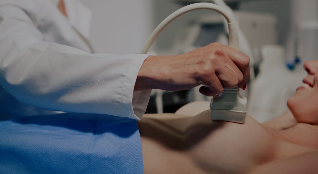
Product Application
Product Features
S-Fetus
Automated Obstetric Ultrasound Work-Flow
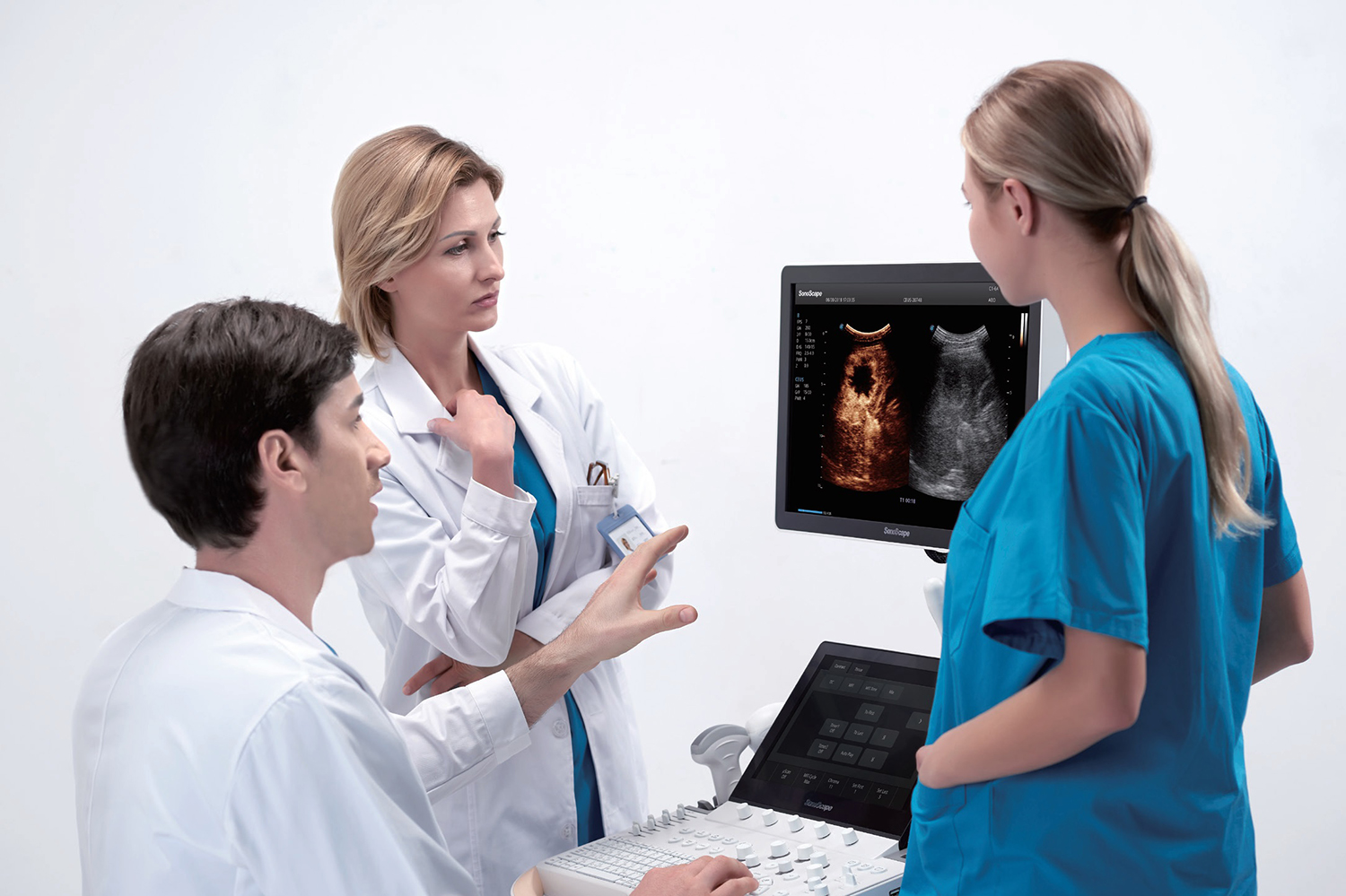
S-Fetus is a simplified function of standard obstetric ultrasound procedures. With just one touch, it can select the best slice image for you, and automatically perform various measurements required to monitor fetal growth and development, transforming obstetric ultrasound examinations into more convenient, faster and more consistent and a more accurate experience.
S-Thyroid
Advanced Tool
S-thyroid is an advanced tool for detecting and classifying suspicious thyroid lesions based on the ACR TI-RADS (American College of Radiology thyroid imaging reporting and data System) guidelines. After selecting the region of interest, s-thyroid can automatically define the boundary of the lesion and generate a report of the characteristics of the suspicious lesion.
Micro F
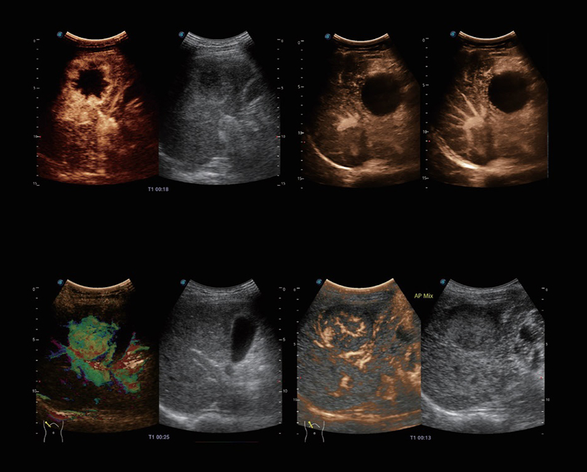
Enables visualization for micro-vascularized structures
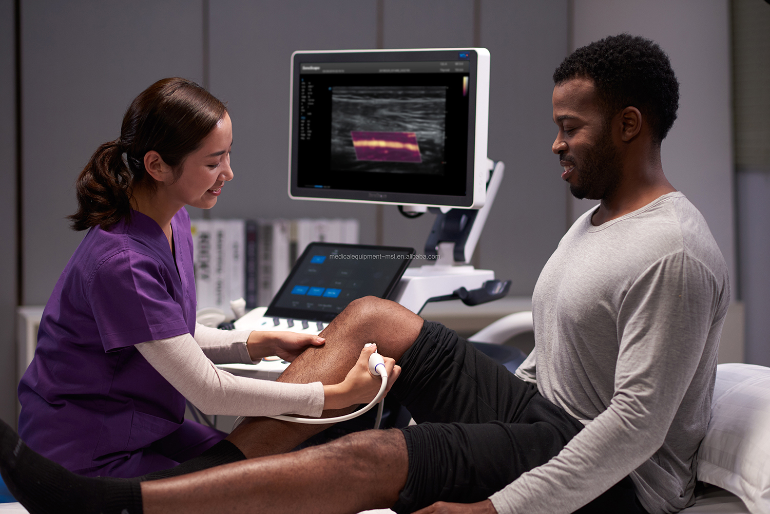
Micro F provides an innovative way to expand the range of ultrasound visible blood flow, especially for the visualization of slowly flowing tiny blood vessels. Micro F uses advanced adaptive filters and the accumulation of time and space signals, which can effectively distinguish tiny flows and covered tissue movements, and describe hemodynamics with higher sensitivity and spatial resolution.
Advanced Cardiovascular
Strives for a comprehensive solution for cardiac evaluation
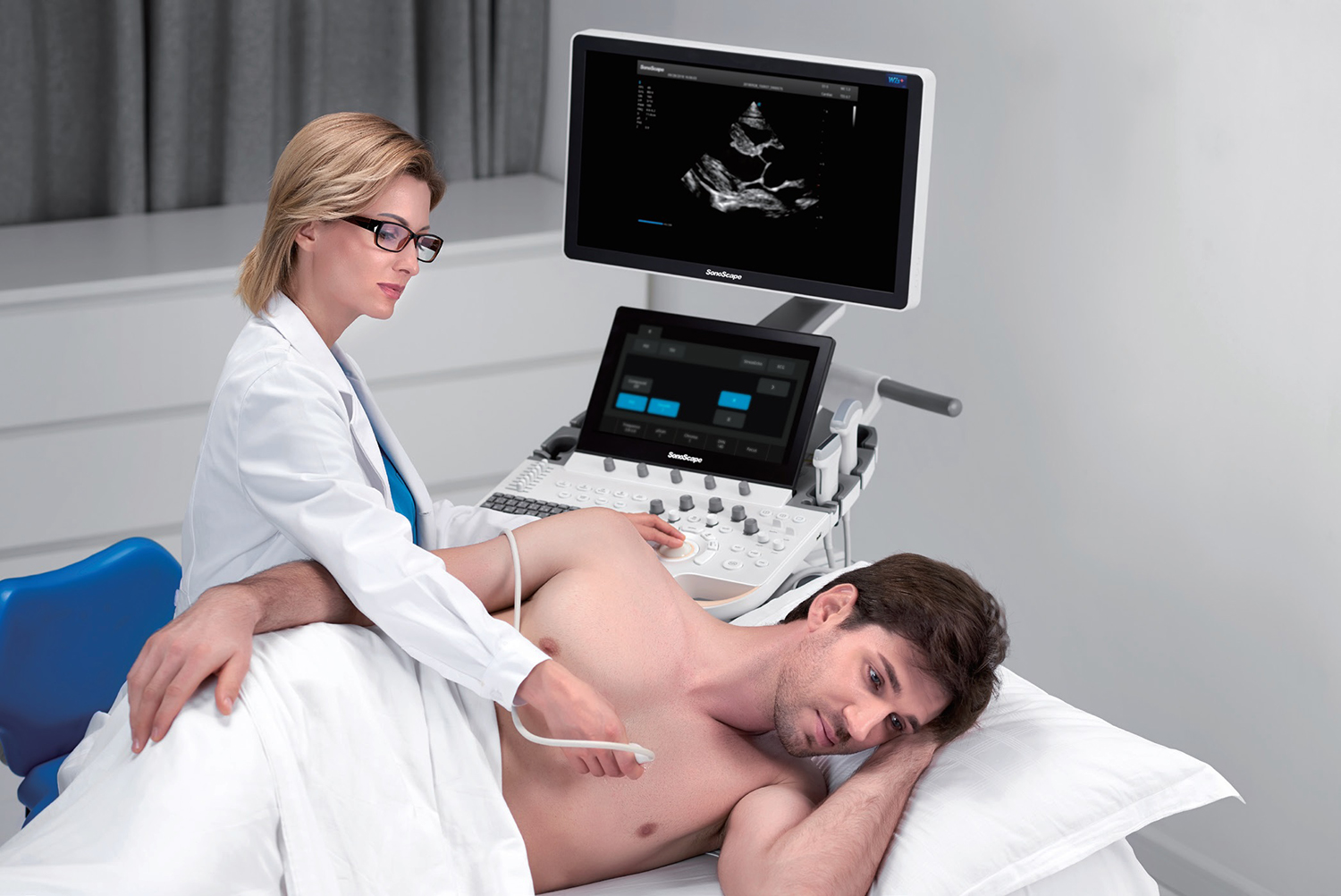
Equipped with SonoScape's unique pure single crystal phased array sensor and the most advanced processing technology, P60 is dedicated to restoring every detail and element to achieve precise diagnosis. New Myocardial Quantitative Analysis (MQA) provides an in-depth quantitative report on the overall and local myocardial wall motion dynamics of the left ventricle, providing doctors with a comprehensive assessment of myocardial function

