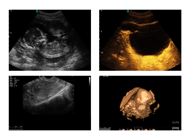
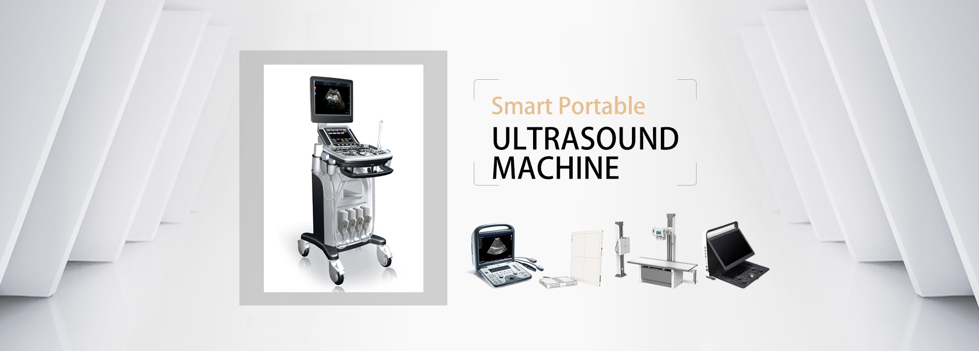
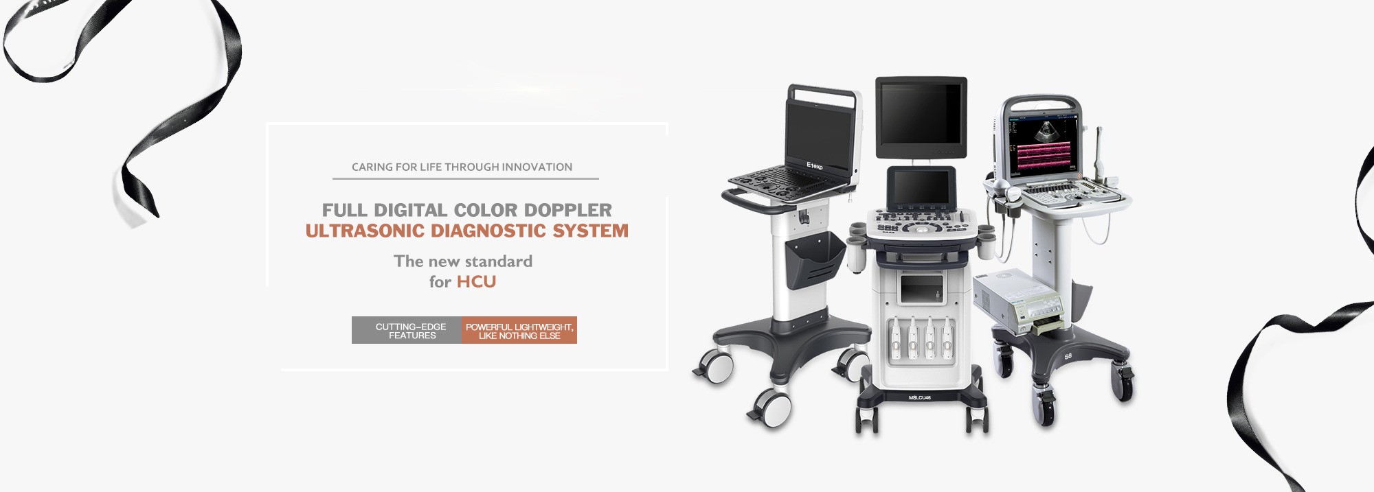
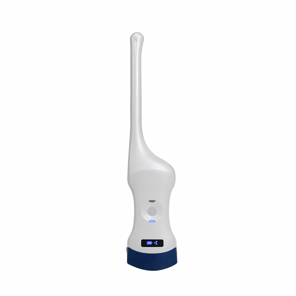
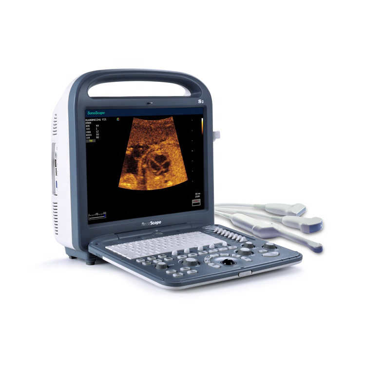
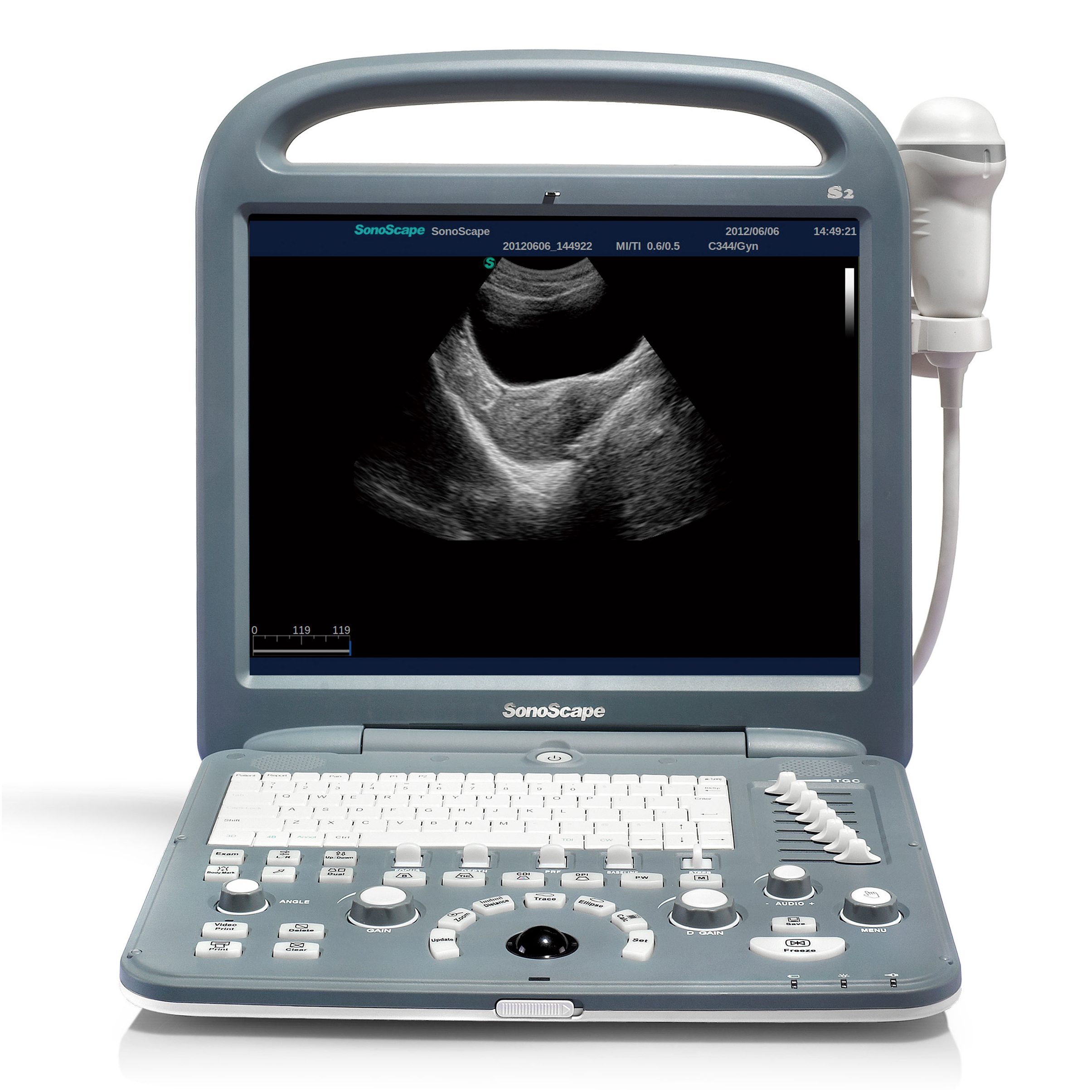
Brief Introduction High-resolution Smart Portable Color Doppler System
Auto Image Optimization
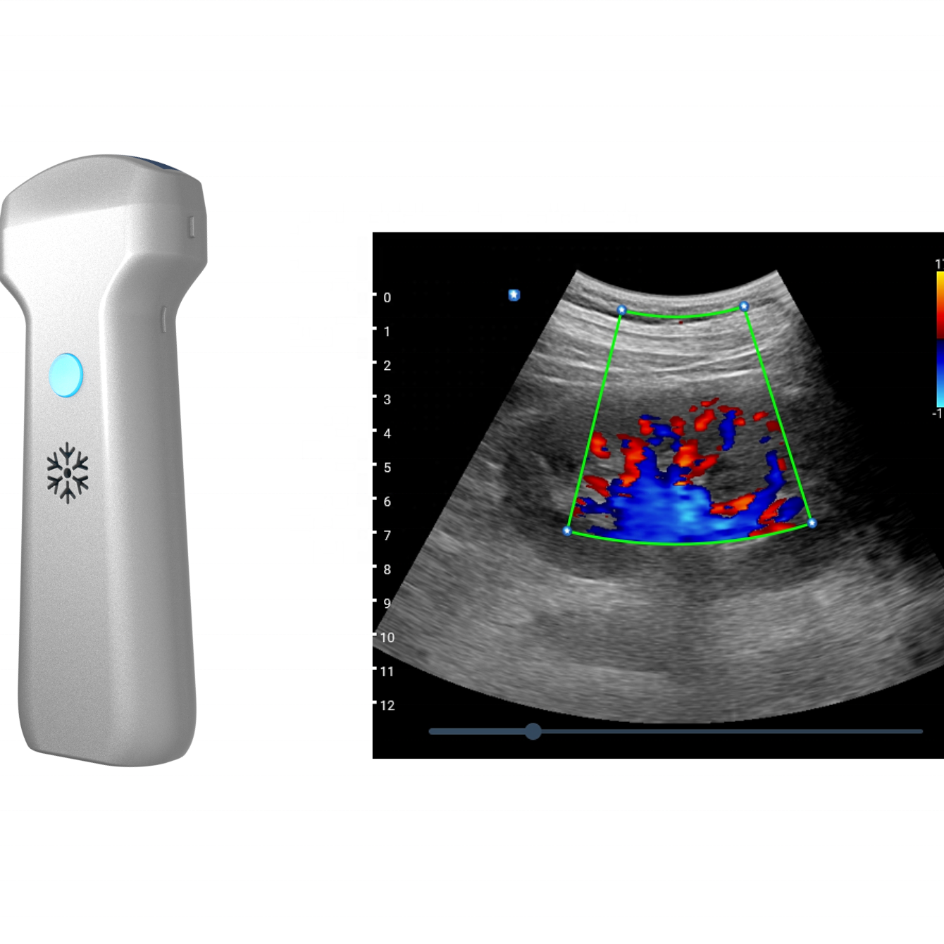
Press a button, the image is automatically adjusted and optimized, saving your time and reducing parameter adjustments. In addition, with Auto Focus on, when the ROI frame moves, the focus area will follow its depth in the scanning area, providing users with ideal image quality of the desired areas.
Automated Calculation
Auto IMT determines the patient's vascular stiffness by automatically tracking the thickness of the carotid artery. Automatic tracking provides users with sensitive and accurate waveform tracking, avoids the error of manual tracking, and gives the calculation results in the first time.

Specifications
|
Scanning Modes
|
B-Mode
M-Mode THI-Mode Color Flow Mode(CFM) Doppler Power Imaging(DPI) PW Mode CW Mode(optional) Panoramic Imaging(optional) 4D Imaging(optional) Steer M-Mode(optional) |
|
Optional Functions
|
4D Imaging
CW Mode Panoramic Imaging IMT Function Anatomic M Mode μ-Scan function |
|
Advanced Technologies
|
Tissue Harmonic Imaging
μ-Scan Speckle Reduction
Compound Imaging
Panoramic imaging
Trapezoid imaging
Freehand 3D and 4D
|
|
General Applications
|
Small Parts(Thyroid, Brest, Testicles and Superficial)
Abdominal(Liver, Spleen, Kidney, Pancreas) Vascular(Carotid, Peripheral vessel) Cardiology Obstetrical(Uterus, Appendages, Fetus) Gynecological Urological Musculoskeletal Craniocerebral Emergency(optional) ICU(optional) Anesthesia (optional) |
|
Sweep Width/Angle
|
Linear Array Maximum 46mm
Convex Array ≥70°
Phased Array ≥90°
Micro Convex Array ≥135°
4D Probe 70°
|
|
Fullfillment of Different Needs
|
Intelligent patient file management system
convenient user-definable settings
Professional diagnostic applications
|
|
Optional Components
|
Power Adaptor
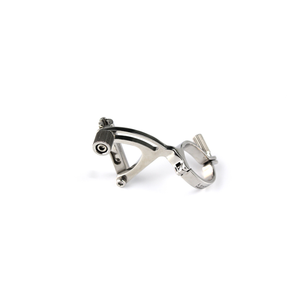
UPS Power Biopsy Guide Color Ink-jet Printer B/W Video Printer External DVD Burner Foot Switch Special Trolley Transducer Cable Holder |


Clinic Images
